
Paperis Articles
The Glymphatic System — Is Sleep When the Brain Rinses Itself?
A 2013 mouse study found the sleeping brain cleared waste faster; a 2024 mouse study found clearance fell during sleep. The live dispute is over what is measured, and how.
Your field's papers, as audio
Pick your fields and journals — Paperis summarizes the key papers and turns them into audio. Listen on your commute.
Start Paperis free →The Glymphatic System — Is Sleep When the Brain Rinses Itself?
Where Does the Brain's Garbage Go?
The brain does not rest even while you sleep. It uses as much energy during REM sleep as it does when awake, and its oxygen consumption drops by only about 20% even in non-REM sleep (PMID 30883420). Working that hard produces byproducts. In other organs, lymphatic vessels collect leftover fluid and debris from between tissues, but there are no such lymphatic vessels inside brain tissue. A lot of garbage is produced, yet the path to clean it away was nowhere to be seen, and this was a long-standing riddle (PMID 30883420).
A candidate for the answer came from the fluid surrounding the brain. The brain and spinal cord are bathed in a clear liquid called cerebrospinal fluid. And the spaces between the cells of brain tissue are filled with a liquid called interstitial fluid. In the early 2010s, a name was given to the idea that these two liquids mix with each other to rinse the brain.
The glymphatic system is a proposed flow in which cerebrospinal fluid enters the brain along microscopic pathways around arteries (perivascular spaces), mixes with interstitial fluid, and carries away waste. The fluid that enters is thought to exit along pathways surrounding veins (PMID 23434588). The name is a combination of glial cells (glia)—the supporting cells of the brain—and lymphatic (PMID 29101220).
In 2013, a research team injected an MRI contrast agent into the space surrounding the mouse spinal cord and imaged it spreading into the brain along the perimeter of the arteries. The research team suggested that this method could one day become a way to gauge the risk of Alzheimer's disease in the living human brain (PMID 23434588). Over the decade since, this flow has been tied to the age-old question "Why Do We Need Sleep?". This article follows how far that pairing is supported and where it is contested. In 2024, an experiment came out showing that cleaning actually decreased in the brain of a sleeping mouse, and in 2025, mechanistic research emerged arguing that sleep promotes the flow. Neither side has received a final verdict yet.
2013, The Brains of Sleeping Mice
When looking inside the brains of living mice and comparing the sleeping state to the awake state, the gaps between cells widened by 60% during natural sleep or under anesthesia, and the flow of cerebrospinal fluid and interstitial fluid mixing through those gaps markedly increased. The experiment also measured the speed at which a protein fragment cleared out. Amyloid-beta is a small protein fragment produced by neurons, and if not cleared away in time, it tangles together and accumulates in clumps outside neurons. This is the clump that accumulates in the brain in Alzheimer's disease. The speed at which this fragment cleared out also became faster while sleeping (PMID 24136970).
A commentary published the same year summarized that cleaning during sleep is up to twice as fast as when awake, and noted that the widening of the gaps may be due to the shrinkage of support cells in the brain called astrocytes. The commentary noted that anesthesia and manipulations that weaken wake-promoting signals can also turn on the same cleaning (PMID 24199995). The authors of the 2013 experiment wrote their conclusion as a conditional statement. They wrote that the reason sleep restores the body and brain may be because it clears away more waste products, which may be toxins accumulated during wakefulness (PMID 24136970).
Mouse results supporting the sleep side continued to appear afterward. In a 2017 experiment, lactate in the brain, which was high during wakefulness, dropped within minutes upon falling asleep or being anesthetized, and suppressing glymphatic function in various ways reduced that drop (PMID 27481936).
What these results brought are two things. One is a new candidate answer to "Why do we sleep?" The other is the observation that anesthesia also alters cleaning in the same direction as sleep. The starting point was a single experiment using mice, and the authors' conclusion was also that "it may be."
The Objections That Were There from the Start
As soon as the hypothesis came out, questions followed. The prevailing view until then was that substances moved through the narrow, tortuous gaps between brain tissue cells mainly by diffusion—that is, spreading naturally from areas of high concentration to low concentration. The glymphatic hypothesis added a directed flow to this, an actual movement of fluid pushed from the arterial side toward the venous side. A 2018 paper wrote that whether this flow was theoretically possible was in doubt, and that the experimental foundation was being challenged by new data (PMID 29101220). A 2020 opinion paper stated that it was expected for the idea of introducing new plumbing to the brain to spark controversy, and neatly sorted what was known, what data was lacking, and what was still being debated (PMID 32423764).
One axis of the debate was the water channel protein embedded in the tips of astrocytes wrapping around blood vessels. The hypothesis viewed this protein as the gateway for the flow, but reports emerged that mice lacking this protein showed no difference in the flow. Five laboratories then set out to re-verify this. The result was that in all four lines with the deleted protein and one line where the protein was missing from around the blood vessels, cerebrospinal fluid entered less than in normal mice, and a meta-analysis combining all the studies also showed a significant reduction in tracer movement (PMID 30561329).
The same paper also accounted for why the results had conflicted. It explained how the type of anesthesia, the age of the animals, and the method of administering the tracer accounted for the opposing results. It also presented findings that inserting a needle directly into the brain tissue suppresses the flow itself, and concluded that future experiments should avoid invasive procedures (PMID 30561329). The lesson left by this dispute surrounding the water channel protein is that how anesthesia is administered and where and what is injected can change the answer. This question does not trap just one side's experiments. A 2017 mouse experiment noted that simply inserting a needle into the cerebrospinal fluid space at the base of the brain (the cisterna magna) suppresses glymphatic function (PMID 27481936), yet subsequent experiments supporting the sleep side also injected tracers into that exact spot (PMID 42367928, PMID 37316849). Invasive procedures and the choice of anesthesia are questions that must be asked equally of experiments on the sleep side and experiments on the opposing side.
2024, Opposite Results
An experiment by a different research team in 2024 reexamined the two states from 2013: sleep and anesthesia. The researchers placed fluorescent molecules in the brains of male mice and measured how much those molecules moved and how fast they left the brain. The movement of the molecules was the same regardless of sleep, waking, or anesthesia. And in the 2024 experiment, brain clearance was clearly reduced during sleep and anesthesia (PMID 38741022).

Illustration: Paperis
This result points in the opposite direction from the 2013 report, using the same animals and the same question. In terms of rating, like the 2013 experiment, this was also a single study done on mice, and the subjects were male. This experiment reported that clearance decreased not only under anesthesia but also during natural sleep, and the point that anesthesia is in the same direction as sleep was the same as in 2013. The point where the two experiments conflict is the direction of clearance: simply put, whether it increases or decreases when you fall asleep. The scope reported by this experiment is up to "the amount of fluorescent molecules leaving the brain decreased during sleep and anesthesia, and molecule movement was independent of state." Even in mice, the proposition that "sleep increases clearance" is disputed over replication.
2025: The Mechanism for the Side That Sleep Pushes the Flow
On the other side stand studies targeting mechanisms. This research focused on non-REM sleep during sleep. Non-REM sleep is the rest of sleep that is not REM sleep, divided into several stages from light sleep to deep sleep. The appearance of the stages is covered in "Sleep Stages and Sleep Cycles". Norepinephrine is a neurotransmitter involved in keeping the brain awake and tense, and it is another protagonist of this study.
A 2025 animal experiment used multiple measurement techniques simultaneously to find that norepinephrine concentration, brain blood volume, and cerebrospinal fluid signals fluctuate slowly in lockstep during non-REM sleep. These three fluctuations most strongly predicted glymphatic clearance. When a cluster of neurons producing norepinephrine was turned on with light, blood vessel constriction and dilation and cerebrospinal fluid signals moved in opposite directions, and when the arteries were made to fluctuate directly, more cerebrospinal fluid entered. The research team interpreted this as evidence that the slow constriction and dilation of blood vessels acts as a pump pushing cerebrospinal fluid into the brain (PMID 39788123). A 2025 review summarizing these studies reported that the rhythm of norepinephrine alters vascular tone and the shape of astrocyte endfeet to control exchange speed, and projected that the breakdown of this rhythm could contribute to the hallmarks of diseases like Alzheimer's disease (PMID 41373600).
In the same experiment, the sleeping pill zolpidem suppressed both the norepinephrine fluctuations and the glymphatic flow (PMID 39788123). This result was measured in animals, and this study did not address what happens in the human brain when a person takes that drug. Separately, in an experiment where young mice had their sleep frequently and chronically disrupted, the slow pulsations of blood vessels weakened, and both glymphatic function and cognitive function declined together. The pulsations created by the heartbeat remained unchanged (PMID 38639025).
This study asked a different question from the 2024 experiment. The norepinephrine research looked at what best predicts clearance and what pushes the flow during non-REM sleep, whereas the 2013 and 2024 experiments compared clearance itself between sleeping and waking states. This study is likewise a single study by one research team. Within the evidence gathered by this article, there is no experiment that compares the two results with the same measurements. The side arguing that sleep pushes the flow also gained new mechanistic evidence in 2025, and that evidence is likewise from animal experiments.
Why They Disagree — What is Measured, and How
Place the three experiments side by side, and the shape of the dispute comes into view. The 2013 experiment measured the speed at which amyloid-beta cleared out in sleeping mice, and that speed increased (PMID 24136970). The 2024 experiment measured the amount of fluorescent molecules escaping from the brain, and that amount decreased (PMID 38741022). Both experiments measured cleaning, used different substances and measurement methods, and the answers were opposite. The 2025 experiment, rather than comparing cleaning amounts, looked for the signals that best predict cleaning and the driving principles of the flow (PMID 39788123). Picture a single mouse's sleeping night: by the 2013 method, amyloid-beta clears out faster; by the 2024 method, fluorescent molecules clear out less; and by the 2025 method, norepinephrine, blood vessels, and cerebrospinal fluid slowly surge together. Which of these three pictures is closer to an actual single night has not yet been judged.
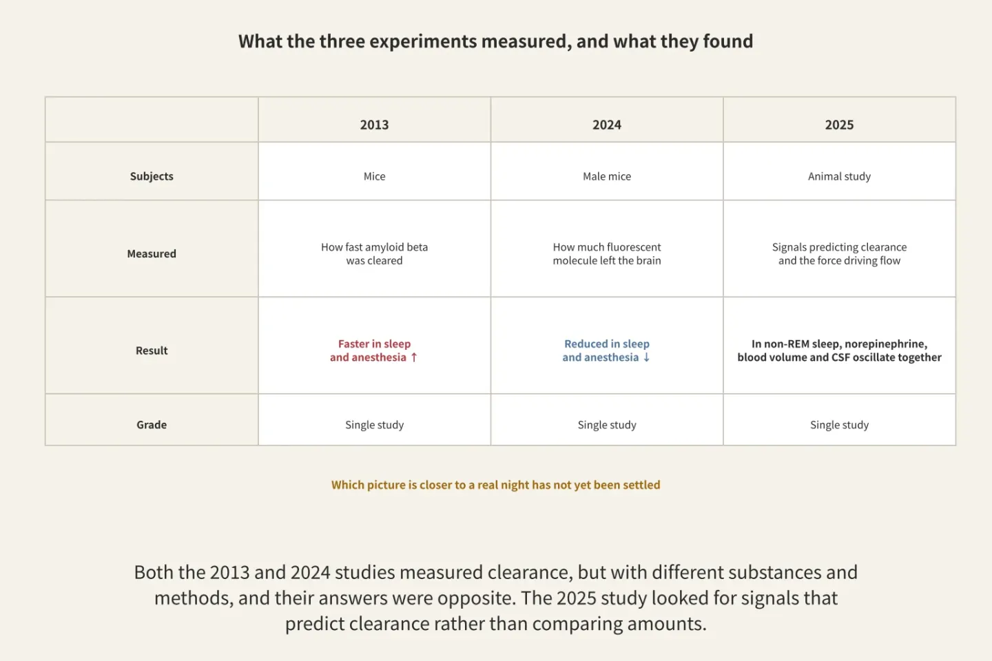
Figure: A diagram drawn by Paperis based solely on the text
Anesthesia is a separate matter to examine. The 2013 and 2024 experiments both measured sleep and anesthesia together, and both reported that anesthesia goes in the same direction as sleep. Since the 2024 experiment reported that cleaning decreased even in natural sleep, the type of anesthesia alone does not explain the discrepancy between the two results. Separate evidence that continues to accumulate is that anesthesia is not a single state. A 2026 preprint (a non-peer-reviewed manuscript) compared two types of anesthesia in mice. In anesthesia using ketamine and dexmedetomidine together, the cerebrospinal fluid entering around blood vessels was much greater than during isoflurane anesthesia. There was an opinion that the lower influx seen in isoflurane might be an optical illusion caused by tracer substances clearing out quickly from the cerebrospinal fluid space at the base of the brain, but even when the same amount was injected slowly over a long time, the difference remained. The authors wrote that this result supports the conclusion that dexmedetomidine-class anesthesia increases glymphatic transport through a mechanism linked to a brain state similar to sleep. This is a conclusion supporting the sleep side hypothesis (PMID 42367928). Another mouse experiment reported that three methods of anesthesia change the pathways through which cerebrospinal fluid escapes in different ways. Isoflurane made the flow exiting toward the nose significantly faster than the other two methods, but decreased the flow exiting toward the spinal cord. This is a small experiment with six mice per group (PMID 41469234). Depending on which exit is measured, the effect of the same anesthesia on cerebrospinal fluid outflow can appear differently.
One mouse experiment separated the variables further. When neural activity and cerebrospinal fluid production were independently controlled using combinations of anesthetic drugs, cleaning efficiency depended on both, and under the condition made similar to sleep, the net exchange ratio was greater than under the condition similar to being awake. In the same experiment, under the awake-like condition with high neural activity, substances had difficulty diffusing into the brain tissue, while cleaning along the perivascular spaces became easier instead. The "sleep-like condition" in this experiment was also a condition created by mixing two anesthetic drugs (PMID 37316849). A 2026 review paper listed as unresolved issues how much flow and diffusion each handle, what differs across animal species, and how to validate human imaging markers (PMID 42419635). The current dispute unfolds over in which state and with what ruler fluid movement is measured.
What is Seen and Not Seen in Humans
The most direct way to see this flow in the human brain is to inject an MRI contrast agent into the space surrounding the spinal cord and take scans again one, two, and four weeks later. When this was done, the contrast agent reached all subregions of the human brain, and compared to a near-healthy baseline group, the clearance speed was slower in a patient group with dementia symptoms caused be normal pressure hydrocephalus. Normal pressure hydrocephalus is a disease caused by blocked cerebrospinal fluid circulation, and it is a different disease from Alzheimer's disease (PMID 29997300). However, this method requires an injection into the spinal canal, so it places a heavy burden on humans (PMID 37655849). A study that fitted computational models to such imaging data from people who had slept and people who stayed up all night reported supporting the idea that diffusion between cells alone is insufficient to explain the movement of tracers in the human brain. The data were explained either by diffusion being roughly 3.5 times faster and separately cleared on the spot, or by flow being added to diffusion. The researchers wrote that the fact that fewer tracers were left behind after an all-nighter is fully explained by a decrease in flow. This was a conclusion obtained from a model, and flow was not directly measured (PMID 37596635).
There is also a small-scale study that asked a different question using the same contrast agent method. Comparing 7 people who stayed up all night and 9 people who slept as much as they wanted, there was no difference in the amount of contrast agent escaping into the sites where lymphatic vessels are located among the membranes wrapping the brain, neither one day nor two days later. The researchers wrote that the data did not support the hypothesis that staying up all night changes this exit (PMID 34587499).
Studies looking at brain pulsations using MRI without contrast agents are even more mixed. In 15 healthy individuals, pulsations in the brain created by blood vessels, respiration, and the heart all increased during non-REM sleep, with blood vessel-induced pulsations increasing the most. The authors viewed this result as supporting the idea that sleep aids fluid movement in the human brain (PMID 35135852). There is also a study that looked for the widening of gaps seen in mice in 2013 within humans. When 21 young individuals were scanned once while awake and once while asleep, the pattern in which water molecules spread changed on the sleep side in the direction expected when extracellular fluid increases. The sleep in this experiment was an all-nighter and sleep induced by sleeping pills, and the authors interpreted this as pointing toward sleep being related to an increase in the volume of extracellular fluid in humans as well (PMID 37084927).
However, in another study, staying up all night increased the slow pulsations created by blood vessels more than non-REM sleep did, while non-REM sleep increased pulsations created by respiration and the heart, with the intensity being greater the deeper the sleep. The authors concluded that the pressure of sleep and deep sleep each engage in the movement of fluid inside the brain in different ways, and perhaps even in waste clearance (PMID 41264592). In a study examining 63 people, the coupling between brain activity and cerebrospinal fluid signals actually became stronger the day after sleep was reduced to 3 hours or less, and this change was seen only in the younger age group. The authors interpreted this as possibly being a compensatory response to a lack of sleep (PMID 40819829). In another study where 36 people were kept awake continuously, the same coupling temporarily strengthened and peaked around the 30-hour mark, and the rhythm of the cerebrospinal fluid signals also temporarily fell out of sync (PMID 41638414). In Stockholm sleep study data (60 people) matching the 63-people study, there was no clear change in the volume fraction of the perivascular space after a single night of sleeping 3 hours or less (PMID 40300399). The degree to which water diffusion inside the brain increased overnight moved in the same direction as the proportion of REM sleep and in the opposite direction as the proportion of non-REM sleep, while the overnight increase in cerebrospinal fluid flow moved with circadian markers regardless of sleep (PMID 34302966).
Let us picture a morning after a poorly slept night. There are two ways to measure amyloid-beta in that person. One is brain imaging. When 20 healthy individuals were kept from sleeping for one night, amyloid-beta burden increased in the right hippocampus and thalamus on PET scans (PMID 29632177). The other is blood. In a 2026 randomized crossover trial where 39 participants alternately experienced normal sleep and an all-nighter, amyloid-beta and tau in plasma were higher on the morning after a normally slept night than after an all-nighter (PMID 41593094). Tau is a protein inside nerve cells that holds the cell's skeleton together, and in Alzheimer's disease, its shape changes and it tangles like skeins of yarn inside the cell. The researchers interpreted this difference as a result of glymphatic clearance increasing during sleep, causing more brain waste to escape into the blood. The authors also suggested that enhancing glymphatic function could become a treatment strategy to slow disease progression. This interpretation relies on nighttime signals measured by an experimental device and a computational model built from published data, and it is a single study involving 39 participants (PMID 41593094).
In neither scene is the flow itself visible. Imaging merely shows the amount left behind in the brain, and blood merely shows the amount that came out into the blood. Broadly speaking, a systematic review combining 51 papers concluded that results between sleep and glymphatic-related markers are mixed in healthy adults. In most studies, there was no correlation between total sleep time and amyloid-beta or tau (PMID 36481512). In humans, flow is being viewed through indirect markers, and those markers sometimes point in different directions.
What a Single MRI Index Can Tell You
The most widely used index in human studies is the ALPS index, obtained via diffusion MRI. It expresses as a single number how much water molecules spread in the direction around blood vessels at a single slice level near the ventricles. Because the calculation is simple and past images can be re-analyzed, it has been widely used for various conditions. A 2024 review paper noted that a high index means water spreads more in that direction at that level, and "nothing more, nothing less." When the index comes out low, the recommendation is not to write "decreased ALPS index" and immediately translate that as "impaired glymphatic function" (PMID 38569866). The problem that values vary across institutions and scanners also remains (PMID 37655849).
In a study comparing 317 people with disrupted sleep and 515 healthy individuals, the ALPS index was lower in the sleep-disrupted group, and the worse the sleep quality score, the lower the index. The authors concluded from this that impaired glymphatic function contributes to disrupting sleep (PMID 37368182). The same correlation can also be read in the opposite direction. A correlation measured at a single time point cannot sort out whether sleep changed the index, or whether something pointed to by the index changed sleep. When you encounter a conclusion written using this index, you can simply keep in mind together that it is a number measuring the spread of water.
The Link with Alzheimer's Disease — Causation in Animals, Correlation in Humans
In animals, the direction of causation can be tested with experiments. In mice where the water channel protein we saw earlier was blocked with a drug, the exchange of cerebrospinal fluid and interstitial fluid decreased, and tau was cleared less. The same study showed the opposite direction as well. In mouse models where tau accumulates, the exchange decreased and the arrangement of that protein was disrupted (PMID 32705145). In another mouse experiment where this protein was blocked for ten weeks, tau tangled more, spread further to the opposite side of the brain, and memory worsened. When the drug was started a few weeks later, those changes were smaller (PMID 38576025). There are also observations matched by age group. Amyloid-beta injected directly into brain tissue was cleared 40% less in old mice than in young mice, the pulsation of small arteries inside the brain decreased by 27%, and that protein was scattered around the blood vessels (PMID 25204284).
In humans, we can only observe. In a study examining the autopsy brains of 79 people, the degree to which the water channel protein was clustered around blood vessels was related to the presence of Alzheimer's disease, regardless of age. The more disrupted that arrangement was, the more amyloid-beta there was, and the higher the stage of the disease. In people whose cognition remained intact even past the age of 85, that arrangement was maintained. The authors concluded that the loss of this arrangement may be a factor that makes the aging brain vulnerable to protein tangles (PMID 27893874). However, whether this abnormal arrangement is a cause that drives the disease or a consequence left behind by the disease is still under debate (PMID 33499944). An imaging study retrospectively analyzing 140 people, combining 99 with Alzheimer's disease, 35 with mild cognitive impairment, and 6 with subjective cognitive decline, reported a relationship where a lower ALPS index meant more amyloid and wrote that "glymphatic dysfunction drives disease progression," which is the conclusion of the authors of the reanalyzed single-timepoint imaging study (PMID 40679601).
The passage of time is also recorded. A 2013 commentary predicted that public health policies helping middle-aged and older adults sleep enough might be useful for preventing Alzheimer's disease (PMID 24199995). Subsequent reviews continue to maintain that hope. A 2026 review summarized that sleep-dependent glymphatic clearance is increasingly recognized as a plausible contributing factor to Alzheimer's disease (PMID 41981905). A critical reappraisal published around the same time in 2025 summarized that knowledge about glymphatic function and Alzheimer's disease comes mainly from clinical data observing correlations and rodent studies. And it is not yet confirmed that glymphatic dysfunction is a cause of Alzheimer's disease in humans (PMID 40638744). The same paper noted that the lack of a clinical imaging method to measure this exchange in the human brain is the barrier preventing that confirmation. A 2026 review also summarized that the direction of causation in humans must be established by future studies following humans over time, and that the lack of a validated non-invasive marker is a major limitation (PMID 42288169). While it was shown through experiments that depriving humans of a single night of sleep changes amyloid-beta markers (PMID 29632177, PMID 41593094), it is not confirmed whether a lack of sleep causes Alzheimer's disease.
Exit — Lymphatic Vessels in the Membranes Wrapping the Brain
Let's follow the path drawn by the hypothesis from start to finish. Cerebrospinal fluid wrapping the surface of the brain enters the brain along channels around arteries (PMID 23434588). According to a 2025 animal experiment, this influx increases when blood vessels slowly constrict and dilate during non-REM sleep (PMID 39788123). The liquid that has entered mixes with interstitial fluid between cells and exits along pathways around veins (PMID 23434588). Substances carried out from deep within the brain are moved to the boundaries of the brain, and it is thought that lymphatic vessels inside the membranes located there send them out toward the body (PMID 40324376). Whether fluid actually flows or merely diffuses in the middle section of this path, between cells, is still a matter of debate (PMID 29101220, PMID 42419635). The point where 2013 and 2024 experiments clashed is also what and how much escapes in this middle section.
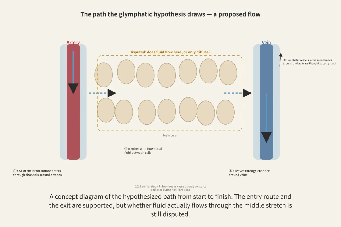
Figure: A schematic drawn by Paperis using only the text of the main body
If there is a flow, there must be an exit at the end. There are no lymphatic vessels inside the brain tissue, but there are lymphatic vessels in the membranes wrapping the brain. In both mice and humans, these membranous lymphatic vessels were placed along large venous passages in the same three-dimensional arrangement, and they are considered pathways that send out waste and handle immune surveillance (PMID 35776089). In a human study looking at 35 patients injected with a contrast agent in the spinal canal, both the clearance of the glymphatic pathway and the clearance of structures presumed to be lymphatic vessels in the membranes slowed with age, and the speeds of the two clearances were interlocked. The researchers viewed this as supporting the idea that the lymphatic vessels in the membranes are the downstream exit of the glymphatic pathway even in humans (PMID 31916277). The all-nighter study seen earlier looked at this very exit side and did not find a difference there (PMID 34587499). The existence of the exit is being supported, but how much sleep changes that exit is still a blank in humans.
What Can Be Said Now
To sum up, three layers remain. On the supported layer, there are these facts. Cerebrospinal fluid enters deep into human brain tissue and flows back out (PMID 29997300), and in mice, a water channel protein wrapping around blood vessels is involved in that movement (PMID 30561329). On the disputed layer, there is the question of whether sleep increases that cleaning. Studies from 2013 and 2025 stand on the side that it increases it, while a study from 2024 stands on the side that it decreases it (PMID 24136970, PMID 39788123, PMID 38741022). Separate evidence has accumulated that the results vary depending on the type of anesthesia (PMID 42367928, PMID 41469234), and that the method of introducing the tracer substance can also change the results (PMID 30561329, PMID 27481936). On the empty layer, there are a way to directly measure the flow in humans and the answer to whether a reduction in that flow causes Alzheimer's disease (PMID 40638744, PMID 42288169).
"Sleep is a time to wash the brain" is a hypothesis opened up by a 2013 mouse experiment, and now it faces opposing results even in the same mice. The places to look next are two. One is a mouse experiment that pits the contradictory results of 2013 and 2024 against each other using the same measurement method, and the other is a validated index that can measure this flow non-invasively in humans.
Source Papers
- "Glymphatic System Function in Relation to Anesthesia and Sleep States." (2019) — PMID 30883420 — Review on the puzzle of the brain's high metabolic rate without lymph vessels in the brain parenchyma, and glymphatic function according to arousal states and anesthesia
- "Brain-wide pathway for waste clearance captured by contrast-enhanced MRI." (2013) — PMID 23434588 — Visualized periarterial influx and cerebrospinal fluid-interstitial fluid exchange using contrast MRI in mice, proposed as a human Alzheimer's evaluation strategy
- "The "glymphatic" mechanism for solute clearance in Alzheimer's disease: game changer or unproven speculation?" (2018) — PMID 29101220 — The conventional view is diffusion; the theoretical validity and experimental foundation of the glymphatic convection hypothesis are challenged
- "Sleep drives metabolite clearance from the adult brain." (2013) — PMID 24136970 — 60% increase in interstitial space, increased clearance of amyloid-beta during sleep and anesthesia in mice, suggesting the restorative function of sleep may be due to waste removal
- "Sleep facilitates clearance of metabolites from the brain: glymphatic function in aging and neurodegenerative diseases." (2013) — PMID 24199995 — 2013 commentary, up to a 2-fold increase in clearance during sleep, outlook that public health sleep policies could be useful for AD prevention
- "Aquaporin-4-dependent glymphatic solute transport in the rodent brain." (2018) — PMID 30561329 — Re-verification by five laboratories, reduced influx in AQP4 deficiency, anesthesia, age, and injection method explain conflicting results, recommendation to avoid invasive procedures
- "Brain clearance is reduced during sleep and anesthesia." (2024) — PMID 38741022 — Male mice, fluorescent molecule movement is independent of state, brain clearance is clearly reduced during sleep and anesthesia
- "Norepinephrine-mediated slow vasomotion drives glymphatic clearance during sleep." (2025) — PMID 39788123 — Synchronized oscillation of norepinephrine, blood volume, and cerebrospinal fluid during NREM sleep is the strongest predictor of clearance, vasomotion acts as a pump, zolpidem suppresses oscillation and flow
- "Chronic sleep fragmentation impairs brain interstitial clearance in young wildtype mice." (2024) — PMID 38639025 — Chronic sleep fragmentation in mice suppresses slow vasomotion and impairs glymphatic function and cognition
- "Enhanced glymphatic CSF tracer influx during α2-adrenergic agonist anesthesia is independent of tracer injection duration." (2026, preprint) — PMID 42367928 — Ketamine/dexmedetomidine shows greater influx than isoflurane, independent of injection duration
- "Anesthetics Modulate Cerebrospinal Fluid Efflux Pathways in Mice by Altering Perineural and Perivascular Spaces." (2026) — PMID 41469234 — Three anesthesia methods alter cerebrospinal fluid efflux pathways differently (6 mice per group)
- "Disentangling the impact of cerebrospinal fluid formation and neuronal activity on solute clearance from the brain." (2023) — PMID 37316849 — Clearance efficiency in mice varies depending on both neuronal activity and cerebrospinal fluid production, net exchange is large under sleep-like conditions
- "The Glymphatic system: A key mechanism linking sleep to brain health and diseases." (2026) — PMID 42419635 — Review citing flow vs. diffusion, species differences, and validation of human imaging metrics as key issues
- "Brain-wide glymphatic enhancement and clearance in humans assessed with MRI." (2018) — PMID 29997300 — Intrathecal contrast agent reaches all subregions of the human brain, delayed clearance in normal pressure hydrocephalus dementia cohort
- "Noninvasive Magnetic Resonance Imaging Measures of Glymphatic System Activity." (2024) — PMID 37655849 — Human tracer methods are invasive; pros and cons of noninvasive MRI metrics and multi-site variability
- "Human brain solute transport quantified by glymphatic MRI-informed biophysics during sleep and sleep deprivation." (2023) — PMID 37596635 — Computational model: diffusion alone is insufficient; reduced clearance after sleep deprivation is explained by decreased advection
- "Cerebrospinal fluid egress to human parasagittal dura and the impact of sleep deprivation." (2021) — PMID 34587499 — 7 all-nighters vs. 9 controls, no difference in dural-side drainage, hypothesis unsupported
- "Human NREM Sleep Promotes Brain-Wide Vasomotor and Respiratory Pulsations." (2022) — PMID 35135852 — 15 people, increased vascular, respiratory, and cardiac pulsations during NREM sleep
- "Sleep deprivation and sleep intensity exert distinct effects on cerebral vasomotion and brain pulsations driven by the respiratory and cardiac cycles." (2025) — PMID 41264592 — All-nighters increase low-frequency pulsations more than NREM sleep does; NREM sleep increases respiratory and cardiac pulsations
- "Increased coupling between global grey matter and CSF-derived fMRI signal in young adults after partial sleep deprivation - evidence from the sleepy brain study" (2025) — PMID 40819829 — 63 people, increased coupling after partial sleep deprivation (young adults only), interpretation of compensatory response
- "Sleep-related and diurnal effects on brain diffusivity and cerebrospinal fluid flow." (2021) — PMID 34302966 — Increase in brain diffusivity overnight is positively correlated with REM ratio and negatively correlated with NREM ratio; increase in cerebrospinal fluid flow is related to circadian rhythm markers
- "The impact of sleep components, quality and patterns on glymphatic system functioning in healthy adults: A systematic review." (2023) — PMID 36481512 — 51 articles, contradictory results for sleep and glymphatic metrics in healthy adults
- "β-Amyloid accumulation in the human brain after one night of sleep deprivation." (2018) — PMID 29632177 — 20 healthy people, increased amyloid-beta burden in the hippocampus and thalamus after one night of sleep deprivation
- "The glymphatic system clears amyloid beta and tau from brain to plasma in humans." (2026) — PMID 41593094 — 39-person randomized crossover trial, morning plasma Aβ and tau are higher after normal sleep, interpretation based on experimental device and multi-compartment model
- "Diffusion Tensor Image Analysis ALong the Perivascular Space (DTI-ALPS): Revisiting the Meaning and Significance of the Method." (2024) — PMID 38569866 — The ALPS index is merely water diffusion in the radial direction at the height of the lateral ventricles, do not use it directly as "glymphatic dysfunction"
- "Glymphatic system impairment in sleep disruption: diffusion tensor image analysis along the perivascular space (DTI-ALPS)." (2023) — PMID 37368182 — 317 sleep disorder patients vs. 515 controls, low ALPS index, authors conclude glymphatic impairment contributes to sleep disorders
- "Impaired glymphatic function and clearance of tau in an Alzheimer's disease model." (2020) — PMID 32705145 — AQP4 inhibition reduces cerebrospinal fluid-interstitial fluid exchange and tau clearance in mice
- "Glymphatic inhibition exacerbates tau propagation in an Alzheimer's disease model." (2024) — PMID 38576025 — 10 weeks of AQP4 inhibition in mice increases tau aggregation and propagation, worsens memory, mitigated when started late
- "Impairment of paravascular clearance pathways in the aging brain." (2014) — PMID 25204284 — 40% reduction in Aβ clearance, 27% reduction in arterial pulsation, loss of AQP4 polarization in old mice
- "Association of Perivascular Localization of Aquaporin-4 With Cognition and Alzheimer Disease in Aging Brains." (2017) — PMID 27893874 — 79 autopsies, loss of perivascular AQP4 arrangement is linked to AD, Aβ, and Braak stages, may be a vulnerability factor
- "Glymphatic system, AQP4, and their implications in Alzheimer's disease." (2021) — PMID 33499944 — Debate ongoing over whether abnormal AQP4 arrangement is a cause or consequence of AD
- "DTI-ALPS index-assessed glymphatic dysfunction mediates Alzheimer's cognitive decline via amyloid-β-dependent pathways: multimodal PET/MRI study." (2025) — PMID 40679601 — 140 retrospective analysis of 99 AD, 35 mild cognitive impairment, 6 subjective cognitive decline, ALPS decreases according to clinical stage, related to Aβ, authors conclude it drives progression
- "Glymphatic dysfunction in Alzheimer's disease: A critical appraisal." (2025) — PMID 40638744 — Knowledge stems primarily from correlational clinical data and rodent studies, causality unconfirmed in humans, lack of clinical imaging methods is a barrier
- "AQP4-mediated glymphatic clearance: Sleep, neurodegeneration, and the translational gap." (2026) — PMID 42288169 — Causality direction in humans must be established with prospective studies, lack of validated noninvasive markers is a major limitation
- "Conserved meningeal lymphatic drainage circuits in mice and humans." (2022) — PMID 35776089 — Meningeal lymphatics with the same three-dimensional arrangement in mice and humans, waste drainage and immune surveillance
- "Impairment of the Glymphatic Pathway and Putative Meningeal Lymphatic Vessels in the Aging Human." (2020) — PMID 31916277 — 35 patients, clearance of the glymphatic pathway and putative meningeal lymphatic vessels declines with age and are correlated with each other
- "Higher order diffusion imaging as a putative index of human sleep-related microstructural changes and glymphatic clearance." (2023) — PMID 37084927 — 21 young adults, decreased diffusion kurtosis in all-nighter + sleep-aid-induced sleep, interpretation related to sleep and increased interstitial fluid volume in humans as well
- "Biphasic adaptation of gBOLD-CSF coupling during sleep deprivation reflects compensatory enhancement and temporal disruption in glymphatic function." (2026) — PMID 41638414 — Coupling during 36 hours of deprivation temporarily increases up around 30 hours, phase misalignment
- "Effects of one-night partial sleep deprivation on perivascular space volume fraction: Findings from the Stockholm Sleepy Brain Study." (2025) — PMID 40300399 — 60 people, no major change in perivascular space volume fraction after one night of partial deprivation
- "Noradrenergic Slow Vasomotion: The Hidden Fluid Pump Linking Sleep, Brain Clearance, and Dementia Pathogenesis." (2025) — PMID 41373600 — Review stating locus coeruleus rhythm changes vascular tone and astrocyte endfoot shape to regulate exchange, rhythm abnormalities may contribute to AD, etc.
- "Sleep-Dependent Clearance of Brain Metabolites via the Glymphatic System: Implications for Alzheimer's Pathophysiology." (2026) — PMID 41981905 — Narrative review stating sleep-dependent glymphatic clearance is increasingly recognized as a plausible contributing factor to AD
- "Glymphatic clearance controls state-dependent changes in brain lactate concentration." (2017) — PMID 27481936 — Brain lactate drops when transitioning to sleep/anesthesia in mice, that drop is attenuated by glymphatic inhibition such as cisterna magna puncture
- "The Brain's Glymphatic System: Current Controversies." (2020) — PMID 32423764 — Opinion paper summarizing debates sparked by the new plumbing concept, what we know, lack of data, and ongoing controversies
- "Glymphatics and meningeal lymphatics unlock the brain-immune code." (2025) — PMID 40324376 — Review stating glymphatics move deep waste to the brain borders and meningeal lymphatics drain it to the periphery
Sources (44)
- Glymphatic System Function in Relation to Anesthesia and Sleep StatesPMID 30883420
- Brain-wide pathway for waste clearance captured by contrast-enhanced MRIPMID 23434588
- ThePMID 29101220
- Sleep drives metabolite clearance from the adult brainPMID 24136970
- Sleep facilitates clearance of metabolites from the brain: glymphatic function in aging and neurodegenerative diseasesPMID 24199995
- Aquaporin-4-dependent glymphatic solute transport in the rodent brainPMID 30561329
- Brain clearance is reduced during sleep and anesthesiaPMID 38741022
- Norepinephrine-mediated slow vasomotion drives glymphatic clearance during sleepPMID 39788123
- Chronic sleep fragmentation impairs brain interstitial clearance in young wildtype micePMID 38639025
- Enhanced glymphatic CSF tracer influx during α2-adrenergic agonist anesthesia is independent of tracer injection durationPMID 42367928
- Anesthetics Modulate Cerebrospinal Fluid Efflux Pathways in Mice by Altering Perineural and Perivascular SpacesPMID 41469234
- Disentangling the impact of cerebrospinal fluid formation and neuronal activity on solute clearance from the brainPMID 37316849
- The Glymphatic system: A key mechanism linking sleep to brain health and diseasesPMID 42419635
- Brain-wide glymphatic enhancement and clearance in humans assessed with MRIPMID 29997300
- Noninvasive Magnetic Resonance Imaging Measures of Glymphatic System ActivityPMID 37655849
- Human brain solute transport quantified by glymphatic MRI-informed biophysics during sleep and sleep deprivationPMID 37596635
- Cerebrospinal fluid egress to human parasagittal dura and the impact of sleep deprivationPMID 34587499
- Human NREM Sleep Promotes Brain-Wide Vasomotor and Respiratory PulsationsPMID 35135852
- Sleep deprivation and sleep intensity exert distinct effects on cerebral vasomotion and brain pulsations driven by the respiratory and cardiac cyclesPMID 41264592
- Increased coupling between global grey matter and CSF-derived fMRI signal in young adults after partial sleep deprivation - evidence from the sleepy brain studyPMID 40819829
- Sleep-related and diurnal effects on brain diffusivity and cerebrospinal fluid flowPMID 34302966
- The impact of sleep components, quality and patterns on glymphatic system functioning in healthy adults: A systematic reviewPMID 36481512
- β-Amyloid accumulation in the human brain after one night of sleep deprivationPMID 29632177
- The glymphatic system clears amyloid beta and tau from brain to plasma in humansPMID 41593094
- Diffusion Tensor Image Analysis ALong the Perivascular Space (DTI-ALPS): Revisiting the Meaning and Significance of the MethodPMID 38569866
- Glymphatic system impairment in sleep disruption: diffusion tensor image analysis along the perivascular space (DTI-ALPS)PMID 37368182
- Impaired glymphatic function and clearance of tau in an Alzheimer's disease modelPMID 32705145
- Glymphatic inhibition exacerbates tau propagation in an Alzheimer's disease modelPMID 38576025
- Impairment of paravascular clearance pathways in the aging brainPMID 25204284
- Association of Perivascular Localization of Aquaporin-4 With Cognition and Alzheimer Disease in Aging BrainsPMID 27893874
- Glymphatic system, AQP4, and their implications in Alzheimer's diseasePMID 33499944
- DTI-ALPS index-assessed glymphatic dysfunction mediates Alzheimer's cognitive decline via amyloid-β-dependent pathways: multimodal PET/MRI studyPMID 40679601
- Glymphatic dysfunction in Alzheimer's disease: A critical appraisalPMID 40638744
- AQP4-mediated glymphatic clearance: Sleep, neurodegeneration, and the translational gapPMID 42288169
- Conserved meningeal lymphatic drainage circuits in mice and humansPMID 35776089
- Impairment of the Glymphatic Pathway and Putative Meningeal Lymphatic Vessels in the Aging HumanPMID 31916277
- Higher order diffusion imaging as a putative index of human sleep-related microstructural changes and glymphatic clearancePMID 37084927
- Biphasic adaptation of gBOLD-CSF coupling during sleep deprivation reflects compensatory enhancement and temporal disruption in glymphatic functionPMID 41638414
- Effects of one-night partial sleep deprivation on perivascular space volume fraction: Findings from the Stockholm Sleepy Brain StudyPMID 40300399
- Noradrenergic Slow Vasomotion: The Hidden Fluid Pump Linking Sleep, Brain Clearance, and Dementia PathogenesisPMID 41373600
- Sleep-Dependent Clearance of Brain Metabolites via the Glymphatic System: Implications for Alzheimer's PathophysiologyPMID 41981905
- Glymphatic clearance controls state-dependent changes in brain lactate concentrationPMID 27481936
- The Brain's Glymphatic System: Current ControversiesPMID 32423764
- Glymphatics and meningeal lymphatics unlock the brain-immune codePMID 40324376