Paperis Articles
What Is a Cell? — A Small Village Inside a Wall, a Village That Splits in Two
We follow the membrane that chooses what comes and goes, the tiny particles that read copies of genetic instructions to build proteins, cells with and without a nucleus, the mitochondria that moved in long ago and stayed, new cells arising from existing cells that divide — and why viruses are not cells.
Your field's papers, as audio
Pick your fields and journals — Paperis summarizes the key papers and turns them into audio. Listen on your commute.
Start Paperis free →What Is a Cell? — A Small Village Inside a Wall, a Village That Splits in Two
A Drop of Blood
If you lightly prick your tip of your finger with a needle, a single red drop wells up. To the eye, it just looks like red water. Yet that drop is densely packed with particles too small to be seen with the eye.
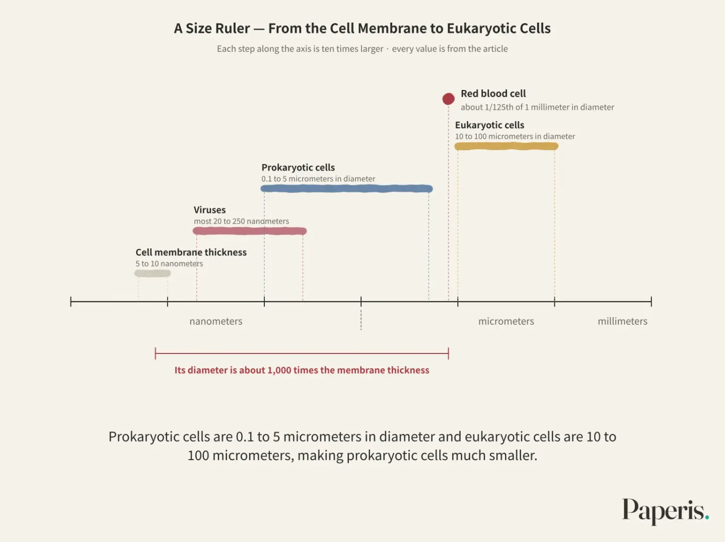
That single particle making the blood red is about 1/125th of 1 millimeter in diameter, so about 250 of them can sit side by side across the width of a pinhead (OpenStax Biology 2e 4.1). The skin on the back of the hand, the leaves outside the window, and the tiny living things that cause disease are all made of such particles. It is just that some living things live as a single one, while others live gathered in countless numbers (OpenStax Biology 2e 4.1).
This article follows that particle, the cell. If you picture a single cell as a small village surrounded by a wall, you will not lose your way. It is a village that separates the inside and outside with a wall, divides work inside the wall, and splits in two when the time comes.
A Name That Came From a Piece of Cork
Cells are invisible to the naked eye except for a few exceptions, so they only came into people's view after the microscope was invented (OpenStax Biology 2e 4.1). The name was given in 1665 from a piece of cork. The scientist Robert Hooke looked through a lens at compartments divided like small rooms and called them cells (OpenStax Biology 2e 4.1). Cells are the smallest units of life, surrounded by a membrane, and new cells arise from cells that already existed. A few years later, the Dutchman Antoni van Leeuwenhoek, who ground lenses himself, observed bacteria (OpenStax Biology 2e 4.1).
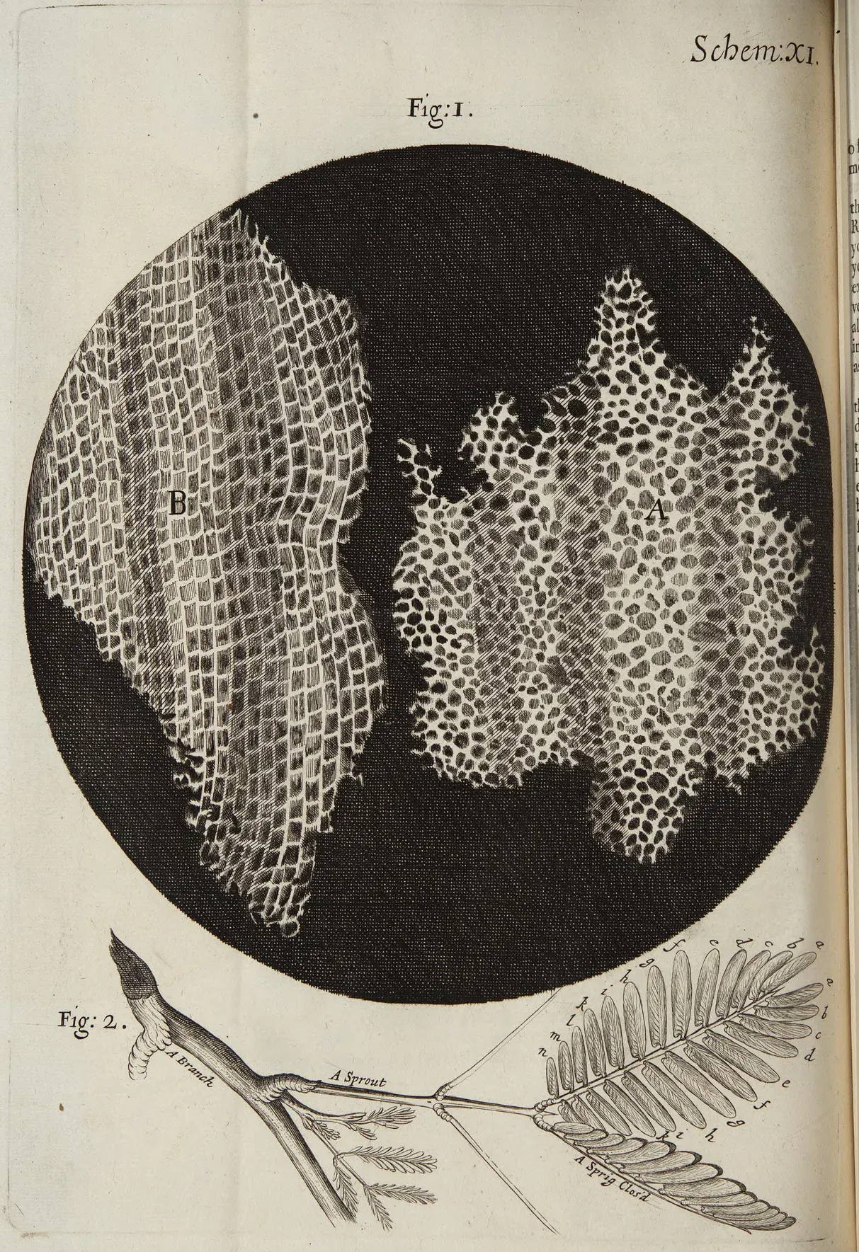
Hooke's engraving of cork cells from Micrographia · Robert Hooke, 1665 · National Library of Wales · Wikimedia Commons (public domain)
Bacteria are tiny living things made of a single cell, and some of them cause disease. Since a single bacterium is a single cell, this means that a single cell alone is enough for a living thing to survive completely (OpenStax Biology 2e 4.1).
In the late 1830s, Matthias Schleiden, who studied plants, and Theodor Schwann, who studied animals, tied these observations together into one idea (OpenStax Biology 2e 4.1). Cell theory is the idea that all living things are made of one or more cells, that the cell is the basic unit of life, and that new cells arise from pre-existing cells. If you want to understand how a living thing works, you just need to look at how a single village runs.
The Wall
There is a wall in a village. The cell membrane is a thin film wrapping a cell, dividing inside from outside, and choosing what goes in and out. Lipid molecules line up in two layers to form the membrane, with proteins embedded between them (OpenStax Biology 2e 4.3). Proteins are molecules made of amino acid chains folded together, and they do most of the work inside the cell. The thickness of the cell membrane is 5 to 10 nanometers, and the diameter of the blood particles we saw earlier is about 1,000 times this membrane thickness (OpenStax Biology 2e 5.1).
Oxygen, water, and nutrients pass through this thin wall, and waste like carbon dioxide goes out (OpenStax Biology 2e 4.3). When the wall collapses, the village ends. A cell that loses the power to select what to let in and what to keep out cannot hold on and breaks apart (OpenStax Biology 2e 5.2).
If we follow where a cell's energy goes, the wall shows up again. Most cells spend most of their energy in two places. One is deliberately keeping the sodium and potassium concentrations different between the inside and the outside of the membrane, and the other is making proteins (OpenStax Biology 2e 5.2). That concentration difference is created by pumps embedded in the membrane. If it is an animal cell membrane, there are pumps everywhere moving sodium and potassium across the membrane, and through that difference, cells regulate their volume and cells like nerves generate electrical signals (PMID 38892309).
There are also signs attached to the wall. Because of markers on the outer surface of the cell membrane, cells recognize each other (OpenStax Biology 2e 5.1). Among the proteins embedded in the membrane, there are also receptors. A receptor is a protein in a cell that recognizes and binds only to specific substances. When a signal like a hormone attaches to this receptor, a reaction continues inside the cell (OpenStax Biology 2e 5.1). The wall is a blocking barrier, and at the same time, a window that receives news from the outside.
Inside the Fence
Even if their shapes differ, almost all cells share four things in common (OpenStax Biology 2e 4.2; OpenStax Biology 2e 22.2). One is the cell membrane we just saw, and the other three are inside the fence.
The cytoplasm is the jelly-like region filling the inside of the cell membrane, where various cell parts float. Even though 70 to 80 percent of it is water, it feels semi-solid because of the proteins dissolved within it (OpenStax Biology 2e 4.3). In a village, it is the ground inside the fence.
Somewhere in that ground lies the manual of the village. DNA is the long molecule where cells store genetic information, and the order in which four bases are lined up is that very information. How that information is written and how it is copied is looked at in detail in "DNA and Genes — A Manual Written in Four Letters, Two Strands That Mirror Each Other".
The manual becomes useful when it is read and made into something. Ribosomes are tiny particles inside the cell that read copies of genetic information and string amino acids together one by one to make proteins. That copy is transcribed from DNA and sent to the ribosome, and is made of RNA (OpenStax Biology 2e 4.3). RNA is a molecule similar to DNA used to transcribe DNA's information or to read that information. The ribosome itself is also a clump of protein and RNA, paired into a large subunit and a small subunit (OpenStax Biology 2e 4.3).
What comes out of this is the very protein embedded in the fence we saw earlier. Proteins make up the largest share of a living body's weight after water, and proteins handle almost all the work that cells do (OpenStax Biology 2e 15.5). A single cell of E. coli, a type of bacterium, contains between 10,000 and 70,000 ribosomes (OpenStax Biology 2e 15.5).
While the detailed parts of ribosomes vary slightly from one organism to another, the overall structure of the protein-making machinery and its job are remarkably similar between bacteria and human cells (OpenStax Biology 2e 15.5). This means that living things with entirely different outward appearances build their workers in the same way. How copies are made and read continues in "From Gene to Protein — A Copy Read Three Letters at a Time, and the Pages Each Cell Opens".
A Village With a Nucleus, A Village Without
When you look at a human cell under a microscope, one lump usually catches your eye first (OpenStax Biology 2e 4.3). The nucleus is a room inside the cell that wraps DNA in a double membrane for safekeeping. The membrane enclosing the nucleus has holes in it, and molecules and RNA travel in and out of the nucleus through those holes (OpenStax Biology 2e 4.3). Like a village, it is as if the instruction manual were kept separately in a house surrounded by a fence.
Cells are broadly split into two groups depending on whether they have a nucleus or not (OpenStax Biology 2e 4.1; OpenStax Biology 2e 4.2).
A prokaryotic cell is a cell that lacks a nucleus and does not have separate rooms enclosed by membranes. A chromosome is a bundle made of long DNA folded and clumped together with proteins. The DNA of a prokaryotic cell is usually a single ring-shaped chromosome, sitting in a zone in the center of the cell. In most prokaryotic cells, a sturdy wall wrapping one more layer outside the cell membrane protects the cell and gives it shape, and some bacteria move with thin thread-like tails (OpenStax Biology 2e 22.2). Organisms made of prokaryotic cells are bacteria and archaea (OpenStax Biology 2e 4.2). Archaea are small single-celled organisms that, like bacteria, have no nucleus, but form a separate lineage from bacteria. Today, living things are divided into three lineages: bacteria, archaea, and organisms made of eukaryotic cells (OpenStax Biology 2e 23.1).
A eukaryotic cell is a cell that has a nucleus and, besides that, is equipped with various rooms enclosed by membranes. Animals, plants, fungi like mushrooms, and protists that fit nowhere else are all made of eukaryotic cells (OpenStax Biology 2e 4.2). An organelle is a small room inside a eukaryotic cell that divides compartments with a membrane and takes charge of a different job. Because the rooms are divided, different jobs can be done separately in different places (OpenStax Biology 2e 4.3). Membrane rooms that trim proteins, wrap them in bundles, and send them off are examples of that (OpenStax Biology 2e 4.4).
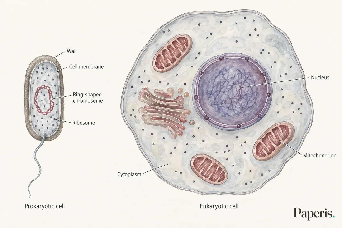
In a small, single-room village, anything that comes in spreads across the whole village instantly, but a large village with many rooms has to pave separate roads to carry goods. Cells are just like that. Prokaryotic cells are 0.1 to 5 micrometers in diameter and eukaryotic cells are 10 to 100 micrometers, making prokaryotic cells much smaller, and inside that small body, incoming substances spread quickly. Large eukaryotic cells are equipped with separate structures to transport substances inside (OpenStax Biology 2e 4.2).
An Old Neighbor That Settled Inside the Fence
One of the rooms in a eukaryotic cell has an unusual origin. That room makes the energy the cell will use. ATP is a molecule that stores the energy a cell is about to use. This is the primary molecule that carries energy around inside the cell (OpenStax Biology 2e 4.3). The mitochondrion is an organelle made of two membranes that uses nutrients and oxygen to make ATP inside a eukaryotic cell. Muscle cells, which use a lot of energy to move the body, have particularly many mitochondria (OpenStax Biology 2e 4.3). It has been revealed over the past half-century that mitochondria take on many more jobs besides ATP, and those jobs vary quite a bit across biological lineages (PMID 29112874).
Mitochondria have their own DNA and ribosomes separately (OpenStax Biology 2e 4.3). Among the people who first saw this small structure with an optical microscope in the late 1800s, a few suggested that a bacterium had come in and was living inside the cell. However, that idea was not accepted for a long time (OpenStax Biology 2e 23.1). In the 1960s, biologist Lynn Margulis at Boston University in the United States refined this idea into a single explanation (OpenStax Biology 2e 23.1).
Endosymbiotic theory is the explanation that the mitochondria of eukaryotic cells came from a bacterium that entered another cell long ago and came to live together. The verdict came down at the molecular level. When comparing the sequences of proteins and RNA between organisms, the comparison of the RNA making up ribosomes in particular gave this explanation the upper hand (PMID 3910132). Today, it is firmly established that mitochondria came from bacteria, and it has even been pointed out which group of bacteria their ancestors were close to (PMID 26439354). This means the room still working inside a human muscle cell is the descendant of an old neighboring village that entered inside the fence long ago and settled down.
What the side that accepted the bacterium—that is, the landlord cell—was remained a major open question even in a 2015 review (PMID 26439354). A 2017 review views that landlord as being close to a group of archaea, but notes that debates continue over the identity of the bacterium that first entered and whether that bacterium settled in before or after other features of eukaryotic cells (PMID 29112874). A 2024 review summarizes that important details about the identities of the landlord cell and the incoming bacterium have been revealed by genome research over the past 10 years (PMID 39261613). Eukaryotic cells are thought to have arisen roughly 1.8 billion to 2.7 billion years ago, but the exact timing and what pushed that event along are still not well understood (PMID 39261613). The mitochondria of eukaryotes are not all the same either. Most have typical mitochondria, but a few leave behind only heavily reduced traces or have no detectable mitochondria (OpenStax Biology 2e 23.1).
The Path of a Single Breath
We follow a single cell in the leg muscles climbing stairs.
Oxygen brought into the body by a breath (OpenStax Biology 2e 7.4) crosses the cell membrane to enter the cell (OpenStax Biology 2e 4.3). On the inner membrane of the mitochondria, electrons from nutrients pass in turn among protein complexes, and at the end of this line, oxygen accepts the electrons to become water (OpenStax Biology 2e 7.4). Most of the ATP produced when burning nutrients using oxygen comes from this process (OpenStax Biology 2e 7.4). This is why muscle cells contain so many mitochondria (OpenStax Biology 2e 4.3).
Carbon dioxide produced as waste from the same work passes back out across the cell membrane, and the carbon dioxide we release with every exhale is precisely what was formed inside cells in this way. When oxygen is in short supply, cells cannot make much ATP (OpenStax Biology 2e 4.3).
This is the path where a single breath passes a fence, is used in an ancient neighbor's room, and crosses back over the fence to leave. At the end of the act of breathing lies the work of cells inside the body taking in oxygen and giving off carbon dioxide (OpenStax Biology 2e 4.3).
Shape Follows Function
Red blood cells, which make blood red, have no nuclei. Red blood cells have no separate structure other than their cell membrane to support the body, and the markers by which other cells recognize them, the properties that let substances in and out, and the properties of bending and withstanding pressure all come from that membrane (PMID 18988878). Cells inside the small intestine that absorb nutrients fold their membranes into finger-like projections to increase their surface area (OpenStax Biology 2e 4.3). Cells in the human body thus vary greatly in shape and interior depending on their assigned tasks, and those shapes are tailored to what they do (OpenStax Biology 2e 4.3).
Those cells lining the inside of the small intestine do not work alone. A tissue is a mass formed by cells that do the same job sticking together. Such tissues gather to become an organ called the small intestine, and organs like the small intestine group and work together to form a single human body (OpenStax Biology 2e 4.1).
How many cells are there in just one such body? This number cannot be counted one by one and is only estimated. At one time, those estimates varied wildly by 10,000-fold, ranging from 1 trillion to 10 quadrillion, and were often cited without a source (PMID 23829164). A 2016 estimate, recalculated by gathering the latest data based on a standard adult weighing 70 kilograms, placed the number of human cells at about 30 trillion, among which about 90% are cells of the blood-forming lineage (PMID 27541692). Another estimate from 2013 added them up by organ to arrive at about 37 trillion (PMID 23829164). Microbes living in the same body are also estimated at about 38 trillion, which is on the same scale as human cells (PMID 27541692).
One Becomes Two
All those many cells are formed by existing cells dividing (OpenStax Biology 2e 10.1). Cell division is the process where a single cell grows, copies its DNA once more, and then splits into two, resulting in two cells with identical DNA. There is also another style of division that makes cells with only half the DNA, like sperm and egg cells, which is covered in "Heredity and Variation — One Card from Each Pair, a Hand Shuffled Anew".
First, the cell grows and copies its DNA. Next, it separates the copied DNA to opposite sides, and finally, the cytoplasm splits to yield two genetically identical cells (OpenStax Biology 2e 10.1; OpenStax Biology 2e 10.2). The DNA of a eukaryotic cell is divided and packed into multiple chromosomes. A human body cell has 46 chromosomes, and sperm and egg cells have half that, 23 (OpenStax Biology 2e 10.1).
Cells are newly made every day. In the human body, 10 billion to 100 billion cells are newly created each day, and an equal number of cells die to maintain the body's balance (PMID 29400998). When the body is normal, dying cells usually die quietly, and immune cells quickly swallow and clear them away (PMID 29400998).
The speed of division varies greatly from cell to cell. In a newly formed embryo, cells divide every few hours, and cells covering the outside and inside of the body are replaced on average every 2 to 5 days (OpenStax Biology 2e 10.3). On the other hand, some cells, like nerve cells in the cerebrum or heart muscle cells, live without ever dividing throughout life (OpenStax Biology 2e 10.3).
When a village gets too large, the length of the wall cannot handle the inner housekeeping. Cells are the same. As their body gets larger, their surface area shrinks compared to their volume, making the housekeeping too burdensome, and that size can be a trigger that calls for division (OpenStax Biology 2e 10.3). The trigger also comes from the outside. When neighboring cells die or hormones that promote growth spread, division can begin, and when cells are crowded closely together, division can be held back (OpenStax Biology 2e 10.3). The driving force pushing cells toward division is a chain of signals turned on by growth-promoting signaling molecules (PMID 36045194).
Neither a Wall nor a Workbench of Its Own
There is a disease that leaves spots on tobacco leaves. Even when the sap of diseased leaves was filtered through a porcelain filtering device that filtered out all bacteria, the filtered sap still transmitted the disease to healthy leaves. This is what researcher Dmitri Ivanovsky showed in 1892 (OpenStax Biology 2e 21.1). There was something much smaller than bacteria. It was only later that its identity turned out to be something completely different from bacteria (OpenStax Biology 2e 21.1).
A virus is a very small pathogen that cannot multiply on its own and must enter the cells of another living thing to multiply. Most are 20 to 250 nanometers in diameter, and except for a few large ones, they cannot be seen with a light microscope (OpenStax Biology 2e 21.1). Viruses are not made of cells. They lack a cell membrane, cell organelles, and ribosomes, having genetic material wrapped in a protein coat, and some viruses wrap the outside of that in an extra layer of membrane obtained from the cell they entered (OpenStax Biology 2e 21.1).
Having neither its own cell membrane nor ribosomes, a virus can multiply only by getting inside someone else's cell. Many viruses grab receptors on the cell surface to attach, and can only multiply inside the cell after piercing the membrane and entering (OpenStax Biology 2e 21.1). Human immunodeficiency virus (HIV) also hijacks receptors on the cell membrane to enter inside the cell (OpenStax Biology 2e 5.1). The genetic material of the virus that has gone inside commandeers the cell's machinery to make copies of itself and its own proteins (OpenStax Biology 2e 21.1). Bacteria get infected by viruses too. From bacteria to plants and animals, there are viruses that infect them across almost every branch of life (OpenStax Biology 2e 21.1).
From the perspective of cell theory, viruses are outside of cells. Various hypotheses have been proposed about where viruses came from, but there is not yet a single unified explanation (OpenStax Biology 2e 21.1).
Where the Village Analogy Cannot Reach
The analogy of a village has its limits. A village is populated by people who think and choose, but the walls, doors, and workbenches of a cell are all molecules, and nothing inside it thinks. If you draw workers at every ribosome or an elected village head commanding the nucleus, the picture becomes wrong. We use the word village only up to the point of bringing to mind the division of inside and outside, rooms and tasks.
Summarized within that scope, it is this. A cell separates inside from outside with a wall and chooses what goes in and out. Inside the wall, an instruction manual called DNA and workbenches called ribosomes make proteins, and if it is a eukaryotic cell, rooms like the nucleus and mitochondria divide the work. A new cell comes to be when a pre-existing cell splits in two, but not all cells have the same shape and not all cells divide.
Where to Look Further
We still do not know for sure exactly when or pushed by what the complex village called a eukaryotic cell came to be (PMID 39261613).
The story of how a single cell sends signals using the electrical difference across its wall can be found in "How Do Nerve Cells Send Signals? — A Burning Fuse, a Gap Crossed by Ferry", and signals read only by receptors on the wall in "How Hormones Work — Letters Only the Addressee Reads, Replies That Say Stop". How cells distinguish invaders from themselves is covered by "How Does the Immune System Tell 'Self' from 'Other'? — Guards at the Gate, and a Patrol That Remembers Faces", and the bacteria living together in the human body by "What Do Gut Microbes Do? — Guests Who Eat the Leftovers, Guests Who Hold the Room". The question of what shapes chains linked together by ribosomes fold into leads on to "From Sequence to Shape: How AlphaFold Cracked a 50-Year Problem".
The other parts of this series open the manuals of this village one by one. "DNA and Genes — A Manual Written in Four Letters, Two Strands That Are Each Other's Molds" looks at how the manual is written, and "From Gene to Protein — A Copy Read Three Letters at a Time, and the Pages Each Cell Opens" at how that manual becomes workers. "Heredity and Variation — One Card from Each Pair, a Hand Shuffled Anew" follows how the manual is mixed and passed down from parents to children, and "What Is Evolution? — Branches Split from One Root, New Shoots Still Sprouting" traces how that manual has changed across generations.
Source Papers
- "Biology 2e — 4.1 Studying Cells" (2018) — OpenStax Biology 2e 4.1 — cells are the smallest units of life, similar cells group into tissues→organs→organ systems, cells are usually invisible to the naked eye with red blood cells at about 8μm, Hooke, Leeuwenhoek, Schleiden, Schwann and cell theory, prokaryotic/eukaryotic two categories.
- "Biology 2e — 4.2 Prokaryotic Cells" (2018) — OpenStax Biology 2e 4.2 — bacteria and archaea are prokaryotes, all cells share four common features (membrane, cytoplasm, DNA, ribosomes), prokaryotic cells 0.1~5μm vs eukaryotic cells 10~100μm and the resulting differences in material movement.
- "Biology 2e — 4.3 Eukaryotic Cells" (2018) — OpenStax Biology 2e 4.3 — eukaryotic cells have a membrane-bound nucleus and organelles, cell membrane structure and transport, cytoplasm water content, nuclear envelope and pores, two-piece ribosomes, mitochondria ATP production, own DNA and ribosomes, muscle cells, small intestine cell villi.
- "Biology 2e — 4.4 The Endomembrane System and Proteins" (2018) — OpenStax Biology 2e 4.4 — the eukaryotic membrane system modifies, packages, and transports proteins and lipids.
- "Biology 2e — 5.1 Components and Structure" (2018) — OpenStax Biology 2e 5.1 — cell membrane markers and receptors, viral receptor hijacking (HIV), membrane thickness 5~10nm making red blood cells about 1,000 times wider.
- "Biology 2e — 5.2 Passive Transport" (2018) — OpenStax Biology 2e 5.2 — cell membranes are selectively permeable and cells cannot survive without this property, most cells use a large share of their energy maintaining sodium-potassium imbalances and synthesizing proteins.
- "Biology 2e — 7.4 Oxidative Phosphorylation" (2018) — OpenStax Biology 2e 7.4 — oxygen is the final acceptor in electron transport along the inner mitochondrial membrane, becoming water, and most ATP from aerobic respiration comes from this process.
- "Biology 2e — 10.1 Cell Division" (2018) — OpenStax Biology 2e 10.1 — the cell cycle produces two genetically identical daughter cells from one cell, human body cells have 46 chromosomes and germ cells have 23.
- "Biology 2e — 10.2 The Cell Cycle" (2018) — OpenStax Biology 2e 10.2 — cells grow and replicate DNA during interphase, divide during the mitotic phase, and become two through cytokinesis.
- "Biology 2e — 10.3 Control of the Cell Cycle" (2018) — OpenStax Biology 2e 10.3 — cell cycle duration ranges from hours in embryos to 2~5 days in epithelia, while nerve and cardiac muscle cells stay in G0 for life, with neighbor cell death, growth hormones, crowding, and size (surface area to volume) regulating division.
- "Biology 2e — 21.1 Viral Evolution, Morphology, and Classification" (2018) — OpenStax Biology 2e 21.1 — filter paper experiments and virus discovery, size 20~250nm, lacking cellular structure (no membranes, organelles, or ribosomes) and requiring attachment to receptors to replicate, viruses exist in almost all life forms, and origin hypotheses remain undecided.
- "Biology 2e — 22.2 Structure of Prokaryotes: Bacteria and Archaea" (2018) — OpenStax Biology 2e 22.2 — four common structures of all cells, prokaryotic circular double-stranded DNA chromosome, most have cell walls and flagella.
- "Biology 2e — 23.1 Eukaryotic Origins" (2018) — OpenStax Biology 2e 23.1 — life has three domains: archaea, bacteria, and eukaryotes; early observations and rejections of mitochondria as bacteria, Lynn Margulis's endosymbiotic theory in the 1960s, and some eukaryotes with reduced or absent mitochondria.
- "Revised Estimates for the Number of Human and Bacteria Cells in the Body." (2016) — PMID 27541692 — recalculates adult human cells at roughly 3.0×10^13 for a 70kg reference man, with about 90% of them belonging to the hematopoietic lineage, and body bacteria numbering roughly 3.8×10^13, on the same scale.
- "An estimation of the number of cells in the human body." (2013) — PMID 23829164 — human cell counts had been cited without sources as 10^12 to 10^16, while summing estimates by organ and cell type yields about 3.72×10^13.
- "Na+/K+-ATPase: More than an Electrogenic Pump." (2024) — PMID 38892309 — the sodium pump sits in the membranes of all animal cells, using ATP energy to transport sodium and potassium to maintain gradients required for volume regulation and electrical excitability.
- "Red cell membrane: past, present, and future." (2008) — PMID 18988878 — enucleated red blood cells rely on their cell membrane as their sole structural component, responsible for all its properties.
- "The Origin and Diversification of Mitochondria." (2017) — PMID 29112874 — mitochondria perform many functions beyond ATP and vary by lineage, originating from a symbiotic alphaproteobacterium and an Asgard archaeal host, with the exact identity of the original symbiont and timing still debated.
- "Endosymbiosis and Eukaryotic Cell Evolution." (2015) — PMID 26439354 — it is established that mitochondria and plastids came from bacteria via symbiosis, with mitochondria tracing to the alphaproteobacteria lineage, while the identity of the host cell remains an open question.
- "The prokaryote-eukaryote interface." (1985) — PMID 3910132 — protein and nucleic acid sequences, especially comparisons of small ribosomal RNAs, settled the debate in favor of the symbiotic origin of eukaryotic cells.
- "The emerging view on the origin and early evolution of eukaryotic cells." (2024) — PMID 39261613 — eukaryotic cells are thought to have evolved from an archaeal ancestor and bacterial symbiont roughly 1.8 billion to 2.7 billion years ago, though the timing and driving forces of complex features remain poorly understood.
- "Apoptosis and Clearance of Apoptotic Cells." (2018) — PMID 29400998 — the human body produces 10 billion to 100 billion cells daily with an equal number dying to maintain homeostasis, and apoptosis is a quiet process with macrophages rapidly engulfing the remains.
- "Biology 2e — 15.5 Ribosomes and Protein Synthesis" (2018) — OpenStax Biology 2e 15.5 — proteins are second only to water in mass and perform almost all functions, E. coli has 10,000 to 70,000 ribosomes per cell, and the synthesis machinery is comparable from bacteria to humans.
- "Cell Cycle Progression and Synchronization: An Overview." (2022) — PMID 36045194 — the driving force pushing cell cycle progression is signaling pathways initiated by growth factors.
Sources (24)
- Biology 2e — 4.1 Studying Cells
- Biology 2e — 4.2 Prokaryotic Cells
- Biology 2e — 4.3 Eukaryotic Cells
- Biology 2e — 4.4 The Endomembrane System and Proteins
- Biology 2e — 5.1 Components and Structure
- Biology 2e — 5.2 Passive Transport
- Biology 2e — 7.4 Oxidative Phosphorylation
- Biology 2e — 10.1 Cell Division
- Biology 2e — 10.2 The Cell Cycle
- Biology 2e — 10.3 Control of the Cell Cycle
- Biology 2e — 21.1 Viral Evolution, Morphology, and Classification
- Biology 2e — 22.2 Structure of Prokaryotes: Bacteria and Archaea
- Biology 2e — 23.1 Eukaryotic Origins
- Revised Estimates for the Number of Human and Bacteria Cells in the BodyPMID 27541692
- An estimation of the number of cells in the human bodyPMID 23829164
- Na+/K+-ATPase: More than an Electrogenic PumpPMID 38892309
- Red cell membrane: past, present, and futurePMID 18988878
- The Origin and Diversification of MitochondriaPMID 29112874
- Endosymbiosis and Eukaryotic Cell EvolutionPMID 26439354
- The prokaryote-eukaryote interfacePMID 3910132
- The emerging view on the origin and early evolution of eukaryotic cellsPMID 39261613
- Apoptosis and Clearance of Apoptotic CellsPMID 29400998
- Biology 2e — 15.5 Ribosomes and Protein Synthesis
- Cell Cycle Progression and Synchronization: An OverviewPMID 36045194