Paperis Articles
Atoms and Radiation — A Drizzle That Never Stops, a Rain Gauge for How Wet We Get
Much of radiation comes from atomic nuclei that have lost their balance. The question is how much we receive — the harm of large doses is clear, and the harm of small doses is still disputed.
Your field's papers, as audio
Pick your fields and journals — Paperis summarizes the key papers and turns them into audio. Listen on your commute.
Start Paperis free →Atoms and Radiation — A Drizzle That Never Stops, a Rain Gauge for How Wet We Get
The Constant Drizzle
When people hear a doctor say, "Let's take a CT scan," many pause first at the word "radiation." Yet that person is receiving radiation even as they sit in the clinic chair. People always live while receiving radiation (PMID 42551708).
This article follows radiation by comparing it to rain. We are all walking in a ceaseless drizzle, and an X-ray or CT scan is a streak of rain added on top for a brief moment. Therefore, the question to ask is not "Did you get hit by rain?" but "How wet did you get?"
That a heavy downpour harms the body is well established. The drizzle side has fewer research studies, and there is a major debate over how harmful it is (PMID 42551708). This article starts from where the raindrops come from, and goes up to the rain gauge that measures the amount of wetting.
The Nearly Empty Atom
When small particles are fired at very thin gold foil, most of the particles pass right through. When the physicist Ernest Rutherford and his colleagues performed this experiment in 1909 using particles from radioactive material, occasionally a particle was deflected at a large angle, and about 1 in every 8,000 bounced straight back toward the source. Almost all of the mass of a gold atom is concentrated in a tiny single point, and the rest is mostly empty space (OpenStax Physics 22.1).
An atom is a particle made up of a small nucleus and electrons surrounding it. The atomic nucleus is a tiny lump in the middle of the atom where almost all of the atom's mass is concentrated.
Electrons are very light, negatively (−) charged particles located in the wide open space around the atomic nucleus.
An atomic nucleus is made of two types of particles. Protons are the positively (+) charged particles that make up the nucleus, and their number determines which element an atom is. Neutrons are the uncharged particles that make up the nucleus, and their mass is nearly equal to that of a proton. Both are much heavier than an electron; a single proton has the mass of 1,836 electrons (OpenStax Physics 22.2).
The name of an element is determined by the number of protons in its nucleus, and different numbers of protons mean different elements (OpenStax Physics 22.1).
Same Element, Different Nuclei
Carbon atoms usually have 6 protons and 6 neutrons. But some carbon atoms have 7 or 8 neutrons, and these are called carbon-13 and carbon-14 (OpenStax Physics 22.2).
Isotopes are atoms of the same element that have different numbers of neutrons in their nuclei, resulting in different masses and stabilities. Hydrogen is a good example. Ordinary hydrogen, deuterium, and tritium behave almost identically chemically, but tritium among them is radioactive (OpenStax Physics 22.2).
The reason a nucleus cannot hold together lies in the tug-of-war inside it. Protons all carry positive electrical charges and push each other apart. That they stick together anyway is because a strong attraction that works only at very close range holds protons and neutrons like glue. This attraction drops almost to zero once it gets just past the size of the atomic nucleus. When the numbers of protons and neutrons do not match and this balance breaks, the nucleus emits something on its own to restore balance (OpenStax Physics 22.2).
Could a stone that never received a single ray of light turn a photographic plate black? In 1896, Becquerel saw that happen. A mineral rich in uranium left a stain on a photographic plate wrapped to keep light from leaking in, and that activity continued without stopping (OpenStax Physics 22.2).
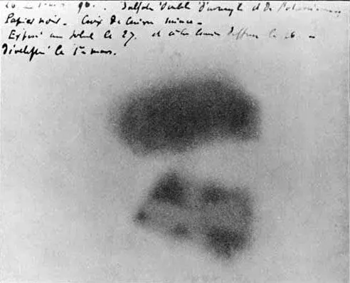
Henri Becquerel's photographic plate fogged by uranium salts · Henri Becquerel, 1896 · Wikimedia Commons (public domain)
Radioactivity is the process in which an atomic nucleus spontaneously emits particles or energy. Radioactive decay is the process in which a nucleus emits something and turns into a nucleus with a different structure. What made Becquerel's photographic plate black was this process (OpenStax Physics 22.2).
What comes out of the nucleus in this way makes up the major branches of radiation. A large share of natural background radiation comes from nuclei that have lost their balance, as does radon, which accounts for roughly half of the amount we receive from nature (PMID 31250572). There is also cosmic radiation flying in from outside the solar system (PMID 18486066). Hospital X-rays come from the electrons inside the atom rather than the nucleus (OpenStax Physics 15.1).
Three Raindrops
There are three kinds of raindrops that come out of an atomic nucleus. Alpha rays are radiation where two protons and two neutrons pop out as a single clump. Because the nucleus loses two protons, the element changes. Uranium gives off alpha rays and turns into thorium (OpenStax Physics 22.2).
Beta rays are electrons that pop out when a neutron turns into a proton inside the atomic nucleus. In this case, the number of protons increases by one, so the element changes as well (OpenStax Physics 22.2).
Gamma rays are electromagnetic waves given off when an excited nucleus settles down into a calm state, and they usually have shorter wavelengths than X-rays. Gamma rays do not change the element (OpenStax Physics 22.2).
In terms of umbrellas, some raindrops are blocked even by a paper umbrella, while others pierce straight through to the roof. A single sheet of paper stops alpha rays, and beta rays are stopped by a thin aluminum plate. Gamma rays require several centimeters of thick lead or thick concrete to be blocked. Alpha rays, carrying two units of electric charge, collide with surrounding atoms right away, whereas gamma rays, having no electric charge, collide less and travel farther. Even within the same kind, if the energy is large they travel farther, and the distance also varies depending on what they encounter (OpenStax Physics 22.2).
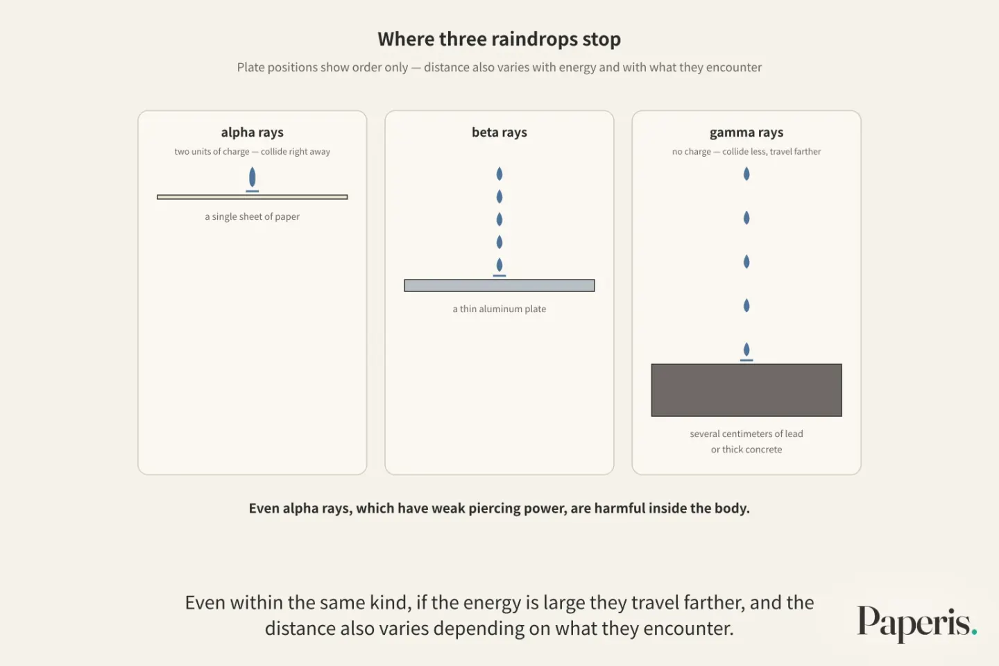
Even alpha rays, which have weak piercing power, are harmful inside the body. Skin usually blocks alpha rays when they come from the outside. However, when they originate inside the body, they concentrate damage over a short distance, making it harder for the body to repair (OpenStax Physics 22.5). An example of decay inside the body is radon. Radon is a natural radioactive gas created when uranium and thorium decay, and when inhaled, it decays inside the lungs and damages them (PMID 31250572).
The Time It Takes for Half to Change
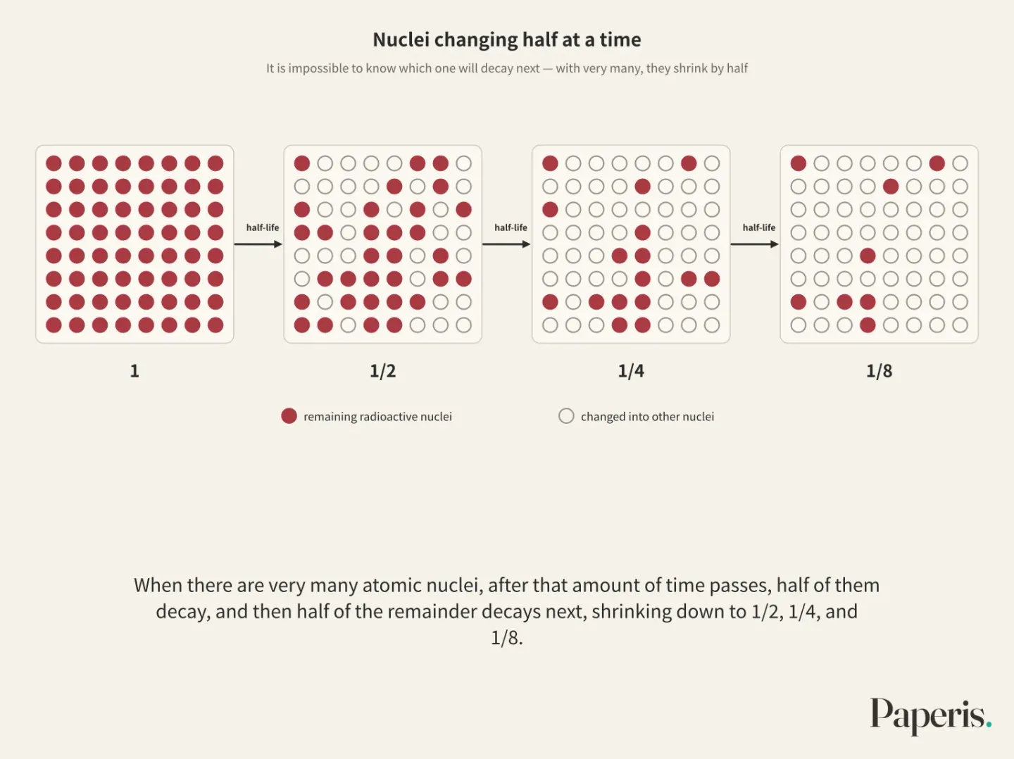
When a very large number of radioactive atomic nuclei are gathered together, it is impossible to know which one will decay next. Each individual nucleus has a 50% probability of surviving for a set amount of time, and no matter how many times it has survived before, the probability for the next identical period of time remains exactly 50% (OpenStax Physics 22.3).
When there are very many atomic nuclei, after that amount of time passes, half of them decay, and then half of the remainder decays next, shrinking down to 1/2, 1/4, and 1/8 (OpenStax Physics 22.3). A half-life is the time it takes for half of a quantity of radioactive atomic nuclei to decay and change into other atomic nuclei. The newly formed atomic nuclei resulting from this change may also be radioactive, which is why radon produced from the decay of uranium causes further harm (PMID 31250572).
Half-lives vary from one atomic nucleus to another. Radium and polonium decay faster than uranium, and correspondingly, more decays take place in the same amount of time (OpenStax Physics 22.3). Among unstable atomic nuclei, some take millions of years to decay on their own (OpenStax Physics 22.4). Measuring the age of ancient objects using half-lives is covered in "Dating with Carbon — Radiocarbon Dates, Read with an Hourglass and Tree Rings".
The Power That Tears Electrons Away
Ionizing radiation is radiation with enough energy to strip electrons from the atoms it hits. Stripping electrons takes very little energy, so even a tiny amount of energy like 0.01 joules causes an immense number of atoms to lose electrons (OpenStax Physics 22.5). Alpha rays, beta rays, gamma rays, and X-rays all belong to ionizing radiation (OpenStax Physics 22.5).
Everything that ionizing radiation does to the body is explained by damage to molecules inside cells, specifically DNA. Yet DNA damage happens all the time even without radiation, up to one million times a day in a single human cell, and cells live by repairing it. Cells whose repair power is broken enter a permanent resting state, kill themselves, or multiply recklessly to become tumors (OpenStax Physics 22.5).
There are places that purposefully gather and use the same rain of rays in one spot. That is cancer treatment. Cancer cells usually divide quickly, and the faster cells divide, the more severely they are hurt by ionizing radiation, so cancer cells collapse before normal cells at the spot where the rays are concentrated (OpenStax Physics 22.5). This is also the path by which cells whose repair power is broken and multiply recklessly, as we saw just before, become cancer, so the same rain of rays spans both the cause and the treatment of cancer.
X-rays and gamma rays are electromagnetic waves, just like radio waves or visible light, and differ only in frequency. Electromagnetic waves with a higher frequency than visible light carry the risk of harming cells, and the likelihood of suffering harm depends greatly on how much you receive (OpenStax Physics 15.1). Both "All electromagnetic waves are dangerous radiation" and "All electromagnetic waves are harmless" are statements made while leaving out the amount and type received. Which side cell phone radio waves fall on, and a map of electromagnetic waves as a whole, are drawn by "Waves — Fans Rising in Their Seats, a Wave Circling the Stadium".
The Markings on a Rain Barrel
Dose is the amount of radiation received by the body or a part of the body, measured by dividing the absorbed energy by the mass of the struck part. Even with the same energy, the dose received by a hand is much greater when concentrated on one hand than when scattered across the whole body, so the dose must always be accompanied by where it was struck (OpenStax Physics 22.5).
Damage differs depending on the type of radiation, even when the same energy is absorbed. Alpha rays act roughly 10 to 20 times more harmfully than X-rays or gamma rays, so when comparing the effects on the body, the absorbed amount is multiplied by a multiplier for each type (OpenStax Physics 22.5).
The numbers appearing in papers mix two things. One is the amount absorbed by a single part of the body, such as the bone marrow, written in milligrays (mGy) (PMID 37946058). The other is the effective dose, written in millisieverts (mSv). The absorbed amount is the energy received per mass by a single part of the body, and the effective dose is a protection value created to compare different scans and exposures on a single scale, making them different scales (PMID 34255191). The effective dose is calculated with a bundle of weights set by the International Commission on Radiological Protection (ICRP), so the value changes depending on which bundle is used (PMID 27893456), and uncertainties and limitations follow (PMID 34255191).
Time is as important as amount. The more amount received across the whole body within a day, the greater the immediate effect, and when the same amount is spread out over a long time, a greater amount is required to produce the same effect (OpenStax Physics 22.5).
The ICRP defines below 100 millisieverts as a low dose. The harm of large doses is well established, and the harm of low doses below 100 millisieverts has little research and is heavily debated (PMID 42551708).
The Amount We Receive
The amount received from nature is estimated at an average of roughly 3.0 millisieverts per year, and among that, 1.8 millisieverts is the share received by inhaling gases called radon and thoron. This is the effective dose value reported by a literature review summarizing the 2024 report of the United Nations Scientific Committee on the Effects of Atomic Radiation (UNSCEAR) (DOI: 10.53862/jupeten.v6i1.004). Because there are also papers writing the amount people receive as about 2 millisieverts a year, like one review article arguing that a threshold should be placed on regulatory standards, the estimates vary quite a bit depending on the source (PMID 40923016).
Just because it comes from nature does not mean it is entirely harmless. Through various epidemiological studies examining miners and general residents, radon has been identified as a cause of lung cancer (PMID 31250572).
In addition to the average 3 millisieverts they receive from nature, people in the United States receive an average of 3 millisieverts more, mainly from medical imaging tests (PMID 31053308). In one survey measuring 12 types of tests, the average effective dose of a single X-ray ranged from 0.01 millisieverts for a side-of-the-head image to 0.5 millisieverts for an abdomen image, and a procedure to widen coronary blood vessels reached 16 millisieverts. The same survey noted that the values for some tests were larger than in existing literature, showing that even the same test yields different values across different surveys (PMID 36516635).
It is reported that long-haul airline pilots receive an average of about 2 to 3 millisieverts per year from flying, and short-haul pilots about 1 to 2 millisieverts. According to this 2008 review, the ICRP recommends a limit for occupational exposure, including flight crews, of an average of 20 millisieverts per year over 5 years, with a maximum of 50 millisieverts in any single year. While noting that epidemiological studies of flight crews did not clearly show an increase in cancer attributable to radiation, the same review judges that there is no threshold below which effects do not occur, only that the probability of suffering harm is very low (PMID 18486066). The amount that nuclear facilities add to their surroundings during normal operation generally stays in the microsievert unit, that is, 1/1,000 of a millisievert (DOI: 10.53862/jupeten.v6i1.004).
The amount received by residents from the 2011 accident at the Fukushima Daiichi Nuclear Power Plant in Japan was estimated by UNSCEAR in a 2013 report, but subsequent studies generally yield lower values than that (PMID 28330389). In nuclear disasters generally, how to prevent early deaths among vulnerable people such as patients already in hospitals during evacuation, and the mental burden on residents living through prolonged shelter life, are cited as tasks that must be addressed alongside radiation regardless of it (PMID 26251394).
The numbers in this section are all effective doses, in millisieverts. Because the milligrays from the CT study appearing later are the amount absorbed by a single site in the bone marrow (PMID 37946058), the two numbers cannot be added together and calculated.
The Harm of Showers Is Clear
For data tracking how harmful radiation is to humans over decades, there is the study of the 1945 atomic bomb survivors in Hiroshima and Nagasaki (PMID 26251394). It follows 93,000 survivors and a comparison group of 27,000 (PMID 22394591).
When a person exposed at age 30 reached age 70, the rate at which solid cancer developed increased by roughly 35% for men and roughly 58% for women above the originally expected rate for every 1 gray of dose received. This does not mean 35% of people get cancer, but rather that the originally expected rate increases by that much. The increased risk appears to persist throughout life (PMID 22394591). The harm of a large dose is this clear.
However, studies analyzing the same data in different ways write it in sieverts, and below 0.2 sieverts, or 200 millisieverts, even with this data, the relationship between the received dose and the risk is murky. This is because in that range, the statistical power to filter it out and the information on the dose each person received are lacking (PMID 24366315).
Does It Get You Wet Even in a Drizzle?
One way to fill in the blur below 200 millisieverts—which even atomic bomb survivor data leaves fuzzy—is to draw a straight line. The linear no-threshold model is a model viewing that the risk of getting cancer from radiation increases in direct proportion to the amount received, and that even the smallest amount carries a risk to that extent. This model has been applied by drawing a straight line from the high risks seen at doses exceeding 1,000 millisieverts down toward lower doses, and it forms the basis of the international radiological protection system (PMID 30907783). It was accepted by multiple countries and international advisory bodies in the 1950s (PMID 30262515).
On one side, evidence is accumulating that risk increases even at low doses. In a study following 948,174 people across 9 European countries who had CT scans before age 22, the higher the cumulative dose received by the bone marrow, the higher the risk of blood cancer. The researchers concluded that this result strengthens the evidence that cancer risk increases at low doses (PMID 37946058). A study following 3.72 million children across 6 US health systems and Ontario, Canada also suggested an association between radiation from medical imaging and a small but meaningfully increased risk of blood cancer (PMID 40961449). A 2021 review summarizes that while evidence that CT scans can cause cancer is growing, the absolute risk to an individual patient is likely small, and adds that brain tumor estimates may have been inflated due to reverse causation (PMID 34545766).
On the other side are reviews arguing that the linear model is wrong. One review points to flaws in epidemiological models. Cancer arises from overlapping causes, and the criticism is that increased cancer was attributed solely to radiation while omitting other risks such as chemicals, diet, and socioeconomic status (PMID 30763555). Another review draws on radiobiological evidence that low doses enhance the body's defenses like DNA repair and immunity, viewing the linear model as highly implausible and models with a threshold or hormesis as plausible. That review also notes that more research is needed on which model fits which situation (PMID 30763552).
Hormesis is the idea that what is harmful in large amounts acts beneficially in small amounts. Reviews criticizing the linear model put forward this idea (PMID 40923016; PMID 30763552). A 2018 review gathering hormesis evidence concludes that the linear model may be outdated and that low doses can be beneficial or ineffective depending on conditions. The same review also introduces studies showing harm at low doses and suggests that because epidemiological studies inherently entail large biases, well-controlled laboratory studies may be more appropriate (PMID 30104556). A 2026 review notes that there is still no consensus regarding the mechanisms of the harmful and beneficial sides, a safe threshold, and long-term outcomes (PMID 42551708).
A 2024 review synthesizing the ground between the two sides judges that low-dose research over the past 10 years is divided among the linear model, models steeper or flatter than the linear model, and the hormesis model, failing to provide a clear answer. And while acknowledging that the linear model plays a part in radiation fear and excessive cleanup costs, it concludes that the evidence to replace it in regulatory models is still insufficient. Whether the risk at low doses is zero, as much as the linear model draws, or greater or smaller than that has not yet been established (PMID 38568156).
A Child's Head CT
When a child falls and hits their head hard, a doctor recommends a head CT. Before the parents' question, the facts can be laid side by side. The numbers given from here are the amount absorbed by the bone marrow, in milligray.
A European study estimated that when children who underwent a CT received an average of 8 milligray to their bone marrow, 1 to 2 out of 10,000 would develop blood cancer within the following 12 years due to radiation (PMID 37946058). To put it the other way around, it means that 9,998 to 9,999 out of 10,000 are estimated not to experience radiation-induced blood cancer during that period. Because this number comes from the overall average of children who underwent CT scans of various body parts, it cannot be directly applied and calculated for a single child's single examination.
Children are more sensitive to radiation than adults and have more years ahead of them to live, making the harm from CT radiation a greater issue for them. At the same time, a carefully weighed and decided CT can have benefits that outweigh the risks (PMID 22472997). A 2014 review addressing CT cancer risk viewed that risk estimate as small, close to background levels, and subject to great uncertainty, and noted that when stating risk with numbers, that uncertainty and the test's benefits must be stated together (PMID 24281269). The same review considers it unjustified to reduce doses so much that it compromises image quality in examinations that are strictly necessary.
European researchers studying CT and brain tumors emphasize carefully weighing whether a child's CT is truly necessary and using doses as low as reasonably achievable (PMID 36493793). In this scene, a small and uncertain risk is placed on one side of the scale, and what can be found out through the examination is placed on the other.
Splitting and Joining Atomic Nuclei
Radiation comes out when atomic nuclei change on their own, but humans can also change atomic nuclei on purpose. Nuclear fission is the event where a heavy atomic nucleus splits into two pieces and releases energy.
When a neutron flies in and embeds itself in a heavy atomic nucleus like uranium, the atomic nucleus elongates like a water droplet and becomes pinched in the middle. Then it splits into two pieces, releasing energy along with a few neutrons. If these neutrons split other atomic nuclei in turn, the fission continues in a tail-chasing manner, which is called a chain reaction (OpenStax Physics 22.4).
Nuclear power plants use the heat obtained by controlling this chain reaction to make steam and generate electricity. In the pressurized water reactor drawn by textbooks, water is needed for the chain reaction to continue, so if the water boils away, the reaction stops on its own, but that is not always enough, and even after it stops, radioactive materials produced by fission generate heat, so a separate cooling device is provided (OpenStax Physics 22.4).
When you sum the masses of the pieces after fission, they fall a little short of the beginning, and the missing mass comes out converted into energy. That amount is the missing mass multiplied by the square of the speed of light (E = mc²) (OpenStax Physics 22.4).
Nuclear fusion is the event where two light atomic nuclei merge into one and release energy. The stumbling block is that both atomic nuclei carry a positive electric charge and push each other away. They must collide fast enough to overcome that repulsion, so even in the sun, nuclear fusion happens only at the center where atomic nuclei are close enough and the temperature is high enough. People generating electricity through nuclear fusion are still at the research stage, and around the time this textbook was written, an international joint test facility was being built (OpenStax Physics 22.4).
The Habit of Asking How Wet We Are
Behind the initial hesitation in front of the CT scanner, there may be two definitive assumptions in opposite directions. One is the thought that "even a tiny bit of radiation is fatal." We are always in a drizzle, cells repair DNA damage that occurs daily, and the body is immediately harmed only when a large dose is received in a short time. The other is the assumption that "low doses are harmless, or even good for the body." Not everything from nature is harmless, as radon is a cause of lung cancer, and large studies following children who underwent CT scans have reported a small but increased risk. Neither the side claiming no harm nor the side claiming it is good for the body has been settled yet (PMID 38568156).
Analogies have limits. Radiation from the outside simply strips away electrons as it passes by and leaves a trace, without pooling in the body like rainwater. However, radioactive substances breathed in like radon decay inside the body (PMID 31250572), and doses received multiple times are added up as cumulative dose to assess the risk (PMID 37946058). Also, even when the same amount falls, the harm differs depending on the type.
The harmfulness of radiation depends greatly on how much is received, and that amount must be read together with the unit, the period, and where on the body. How harmful a single drop of drizzle is has not yet been determined (PMID 38568156).
Where to Read More
- How to date ancient objects using half-life is covered in "Dating with Carbon — Radiocarbon Dates, Read with an Hourglass and Tree Rings".
- The benefits and limitations of screening for lung cancer using low-dose CT are covered in "Lung Cancer and Low-Dose CT Screening — Reading a Small Spot in the Picture Over Time".
- The division of cells into two can be seen in "What Is a Cell? — A Small Village Inside a Wall, a Village That Splits in Two". This forms the basis of why cells that divide quickly are vulnerable to radiation.
- "Waves — Fans Rising in Their Seats, a Wave Circling the Stadium" covers the entire electromagnetic spectrum, and "Energy — Money That Doesn't Shrink When Exchanged, Odd Coins Scattered in Your Pockets" covers energy conservation and conversion.
Source Papers
- "Physics — 22.1 The Structure of the Atom" (2020) — OpenStax Physics 22.1 — Rutherford's gold foil experiment (most pass through, 1 in 8,000 deflects back), mass concentrated in a small nucleus, atomic number (proton number) varies by element.
- "Physics — 22.2 Nuclear Forces and Radioactivity" (2020) — OpenStax Physics 22.2 — Atoms have sub-structure, protons and neutrons, isotopes differ in mass and stability due to neutron number differences, balance between electrical repulsion and the strong nuclear force, Becquerel's discovery, alpha, beta, and gamma decay, penetration distance depends on type, energy, and material.
- "Physics — 22.3 Half Life and Radiometric Dating" (2020) — OpenStax Physics 22.3 — Half-life is the time for half to decay, survival probability of a single nucleus is independent of its past, decay speed varies by nucleus.
- "Physics — 22.4 Nuclear Fission and Fusion" (2020) — OpenStax Physics 22.4 — Neutron-induced nuclear fission (water-drop model), chain reactions, pressurized water reactor water and control rods, decay heat, mass-energy equivalence, nuclear fusion of nuclei lighter than iron and its hurdles, solar core, nuclear fusion test facilities under construction at the time of writing.
- "Physics — 22.5 Medical Applications of Radioactivity: Diagnostic Imaging and Radiation" (2020) — OpenStax Physics 22.5 — Action of ionizing radiation such as X-rays, gamma, alpha, beta explained by DNA damage, cellular DNA repair, large impact on rapidly dividing cells (treatment and cause), dose is absorbed energy divided by mass, multiplier of biological effects by type (approximate value), short-duration high dose and immediate effects.
- "Physics — 15.1 The Electromagnetic Spectrum" (2020) — OpenStax Physics 15.1 — Electromagnetic waves are the same kind with only different frequencies, X-rays come from inner atomic electron transitions, cell damage risk at frequencies higher than visible light heavily depends on the received amount.
- "Low-dose ionizing radiation and cognitive function: evidence, mechanisms, and controversies." (2026) — PMID 42551708 — People are constantly exposed to ionizing radiation, effects of large doses are established, low doses below 100mSv (ICRP definition) are limitedly studied, controversial, and lack consensus on mechanisms, safety thresholds, and long-term outcomes.
- "Health Effects of Radon Exposure." (2019) — PMID 31250572 — Radon is produced by the decay of uranium and thorium and accounts for about half of natural background radiation, when inhaled it decays inside the lungs and damages them, proven cause of lung cancer.
- "Cosmic radiation in commercial aviation." (2008) — PMID 18486066 — Cosmic radiation comes from outside the solar system and varies with altitude, latitude, and solar cycle, long-haul pilots receive 2–3 mSv/year and short-haul 1–2 mSv/year, ICRP occupational limit is a 5-year average of 20 mSv/year (maximum 50 mSv in a single year), no decisive evidence of increased cancer in crew epidemiology, no threshold but probability of harm is very low.
- "Medical Imaging and Pediatric and Adolescent Hematologic Cancer Risk." (2025) — PMID 40961449 — 3.72 million children in the US and Ontario, suggesting an association between medical imaging radiation and a small but significant increased risk of hematological cancers.
- "Risk of hematological malignancies from CT radiation exposure in children, adolescents and young adults." (2023) — PMID 37946058 — 948,174 people across 9 European countries, bone marrow absorbed dose estimated, cumulative dose associated with hematological cancer risk, 1 to 2 people per 10,000 children within 12 years at an average of 8 mGy, strengthening evidence for low-dose risk.
- "Dosimetric quantities and effective dose in medical imaging: a summary for medical doctors." (2021) — PMID 34255191 — Effective dose is a protection quantity comparing doses from different exams and imaging modalities, with uncertainties and limitations.
- "Updated effective doses in radiology." (2016) — PMID 27893456 — Effective dose per examination varies depending on ICRP60 and ICRP103 weighting sets.
- "Bridging Radiation Risk Perception in Indonesia: A Literature Study Comparing Natural and Artificial Radiation Sources Based on UNSCEAR 2024" (2026) — DOI: 10.53862/jupeten.v6i1.004 — Based on UNSCEAR 2024, annual average natural effective dose is about 3.0 mSv (1.8 mSv from radon and thoron inhalation), routine exposure from nuclear facilities is in the µSv range.
- "Radiation in Spine Deformity: State-of-the-Art Reviews." (2019) — PMID 31053308 — Americans receive 3 mSv of natural radiation plus an additional 3 mSv mainly from medical imaging.
- "Questioning the Linear No-Threshold Model (LNT): Lessons From Hiroshima/Nagasaki and Fukushima." (2025) — PMID 40923016 — Lecture and review opposing the linear model and supporting hormesis, noting people receive about 2 mSv annually.
- "Typical effective dose values from diagnostic and interventional radiology." (2023) — PMID 36516635 — Survey of effective doses for 12 exams: general X-ray 0.01 (side of head) to 0.5 mSv (abdomen), mammography 0.5 mSv, coronary angiography up to 16 mSv, some exams larger than literature.
- "Radiation Doses and Associated Risk From the Fukushima Nuclear Accident." (2017) — PMID 28330389 — Recent studies suggest lower doses than the UNSCEAR 2013 report, and the report also noted potential overestimations such as unreflected decontamination.
- "Nuclear disasters and health: lessons learned, challenges, and proposals." (2015) — PMID 26251394 — Nuclear disasters like the 1945 atomic bombs and power plant accidents leave health issues unrelated to radiation, with challenges including preventing premature death of vulnerable groups during evacuation and the psychological burden on long-term evacuees.
- "Radiation and cancer risk in atomic-bomb survivors." (2012) — PMID 22394591 — Follow-up of 93,000 atomic bomb survivors and 27,000 controls over decades, solid cancer incidence at age 70 for exposure at age 30 increases by 35% for men and 58% for women per Gy, risk persists for life.
- "Cancer risk at low doses of ionizing radiation: artificial neural networks inference from atomic bomb survivors." (2014) — PMID 24366315 — Risk below 0.2 Sv is not well defined due to limitations in statistical power and dose information (threshold-side analysis results from this study itself are not used in the main text).
- "Radiation Biology and Its Role in the Canadian Radiation Protection Framework." (2019) — PMID 30907783 — The linear model assumes risk is proportional to dose and has been applied by extrapolating linearly from high doses exceeding 1,000 mSv, forming the basis of the international protection system.
- "Are We Approaching the End of the Linear No-Threshold Era?" (2018) — PMID 30262515 — The linear model was adopted by domestic and international advisory bodies in the 1950s, and the author argues for hormesis and calls for its abandonment, citing atomic bomb survivors, background radiation, and occupational exposure.
- "Epidemiological studies of CT scans and cancer risk: the state of the science." (2021) — PMID 34545766 — Evidence that CT can cause cancer is growing, but the absolute risk for individual patients is likely small, brain tumor estimates may be overestimated due to reverse causality.
- "Ionizing radiations epidemiology does not support the LNT model." (2019) — PMID 30763555 — Criticism that the linear model omits other risk factors like chemicals, diet, and socioeconomics, linearly interpolates high-dose data, and assumes identical mechanisms.
- "The LNT model for cancer induction is not supported by radiobiological data." (2019) — PMID 30763552 — Based on radiobiological evidence, the linear model is highly implausible while threshold and hormesis models are plausible, and more research is needed on which model is correct and when.
- "Overview of Biological, Epidemiological, and Clinical Evidence of Radiation Hormesis." (2018) — PMID 30104556 — Hormesis-leaning review: the linear model may be outdated and low doses can be beneficial or ineffective depending on conditions, introducing harmful studies as well and arguing that controlled laboratory studies are more appropriate due to epidemiological biases.
- "A Review of Recent Low-dose Research and Recommendations for Moving Forward." (2024) — PMID 38568156 — Literature from the past decade is divided among linear, steeper-than-linear, sublinear, or hormetic models with no clear answer, and while the linear model contributes to radiation fear, there is insufficient evidence to change regulatory models.
- "Radiation dose features and solid cancer induction in pediatric computed tomography." (2012) — PMID 22472997 — Children are more sensitive to radiation and have a longer remaining life expectancy making CT risk important, but well-justified CT benefits can outweigh risks.
- "Current knowledge on tumour induction by computed tomography should be carefully used." (2014) — PMID 24281269 — Tumor induction probability estimates from CT are small close to background levels with large uncertainties, predictions should be communicated with uncertainties and benefits, and dose reduction that compromises the image quality of necessary exams is unjustified.
- "Brain cancer after radiation exposure from CT examinations of children and young adults: results from the EPI-CT cohort study." (2023) — PMID 36493793 — Significant dose-response between CT brain dose and brain cancer, emphasizing careful justification of children's CT and doses as low as reasonably achievable.
Sources (31)
- Physics — 22.1 The Structure of the Atom
- Physics — 22.2 Nuclear Forces and Radioactivity
- Physics — 22.3 Half Life and Radiometric Dating
- Physics — 22.4 Nuclear Fission and Fusion
- Physics — 22.5 Medical Applications of Radioactivity: Diagnostic Imaging and Radiation
- Physics — 15.1 The Electromagnetic Spectrum
- Low-dose ionizing radiation and cognitive function: evidence, mechanisms, and controversiesPMID 42551708
- Health Effects of Radon ExposurePMID 31250572
- Cosmic radiation in commercial aviationPMID 18486066
- Medical Imaging and Pediatric and Adolescent Hematologic Cancer RiskPMID 40961449
- Risk of hematological malignancies from CT radiation exposure in children, adolescents and young adultsPMID 37946058
- Dosimetric quantities and effective dose in medical imaging: a summary for medical doctorsPMID 34255191
- Updated effective doses in radiologyPMID 27893456
- Bridging Radiation Risk Perception in Indonesia: A Literature Study Comparing Natural and Artificial Radiation Sources Based on UNSCEAR 2024DOI 10.53862/jupeten.v6i1.004
- Radiation in Spine Deformity: State-of-the-Art ReviewsPMID 31053308
- Questioning the Linear No-Threshold Model (LNT): Lessons From Hiroshima/Nagasaki and FukushimaPMID 40923016
- Typical effective dose values from diagnostic and interventional radiologyPMID 36516635
- Radiation Doses and Associated Risk From the Fukushima Nuclear AccidentPMID 28330389
- Nuclear disasters and health: lessons learned, challenges, and proposalsPMID 26251394
- Radiation and cancer risk in atomic-bomb survivorsPMID 22394591
- Cancer risk at low doses of ionizing radiation: artificial neural networks inference from atomic bomb survivorsPMID 24366315
- Radiation Biology and Its Role in the Canadian Radiation Protection FrameworkPMID 30907783
- Are We Approaching the End of the Linear No-Threshold Era?PMID 30262515
- Epidemiological studies of CT scans and cancer risk: the state of the sciencePMID 34545766
- Ionizing radiations epidemiology does not support the LNT modelPMID 30763555
- The LNT model for cancer induction is not supported by radiobiological dataPMID 30763552
- Overview of Biological, Epidemiological, and Clinical Evidence of Radiation HormesisPMID 30104556
- A Review of Recent Low-dose Research and Recommendations for Moving ForwardPMID 38568156
- Radiation dose features and solid cancer induction in pediatric computed tomographyPMID 22472997
- Current knowledge on tumour induction by computed tomography should be carefully usedPMID 24281269
- Brain cancer after radiation exposure from CT examinations of children and young adults: results from the EPI-CT cohort studyPMID 36493793