
Paperis Articles
The 2026 Nobel Prize in Physiology or Medicine — A Door Opened by Light, From Pond Algae to a Switch for the Brain
The 2026 Nobel Prize in Physiology or Medicine. A question about why eyeless, brainless single-celled algae swim toward light yielded an ion channel that opens by light; its gene, placed in neurons, became a millisecond switch. In people it has so far been used in one organ — the eye — in reports of one patient and of four.
Your field's papers, as audio
Pick your fields and journals — Paperis summarizes the key papers and turns them into audio. Listen on your commute.
Start Paperis free →The 2026 Nobel Prize in Physiology or Medicine — A Door Opened by Light, From Pond Algae to a Switch for the Brain
How Do Green Particles in a Pond Find the Light?
If you draw a drop of pond water, place it on a glass slide, and shine a light from only one side, green particles invisible to the naked eye crowd toward the lit side. They are green algae living as single cells. With no eyes, no brain, and no nerves, they figure out which direction the light is coming from and paddle their tails to go there. In front of light that is too strong, they instead turn their bodies around to avoid it.
To this organism, light is food. Because it receives light and makes its own nutrients, figuring out where it is bright is a matter of making a living. In front of light that is too strong, the opposite reaction of avoiding it appears. Therefore, two reactions together are necessary: seeking out the bright side and avoiding the excessively bright side.
The problem lay in the fact that there was no apparatus in sight to pull off that job. A single-celled organism has no organ called an eye and no nerves to gather signals and make judgments. So the distance between receiving light and changing swimming direction must be very short. The fact that this reaction of swimming toward light and avoiding strong light happens because of a protein that takes in light has been known from early on (PMID 12089443). The remaining questions were what that protein was and how it worked. Research that started from this question, which sought to explain the swimming direction of pond algae, gave birth to the fundamental tools of brain research today.
On October 5, 2026, the Nobel Assembly at the Karolinska Institute in Sweden announced that it would award the Nobel Prize in Physiology or Medicine to three people: Karl Deisseroth of the Howard Hughes Medical Institute and Stanford University in the United States, Peter Hegemann of Humboldt University of Berlin in Germany, and Georg Nagel of University of Würzburg in Germany (Nobel Committee, press release). The reason for the award is the discovery of a protein that responds to light in green algae and turning it into a tool that controls brain cell activity with light. The Nobel Committee explains that Hegemann and Nagel discovered this protein in single-celled algae, and Deisseroth turned that protein into a light switch that turns brain cells on and off (Nobel Committee, press release).
This alga has been given the name Chlamydomonas reinhardtii. It is an organism that commonly lives in ponds and soil, and it is an organism widely used in laboratories. The story begins in a place that has nothing to do with the human brain.
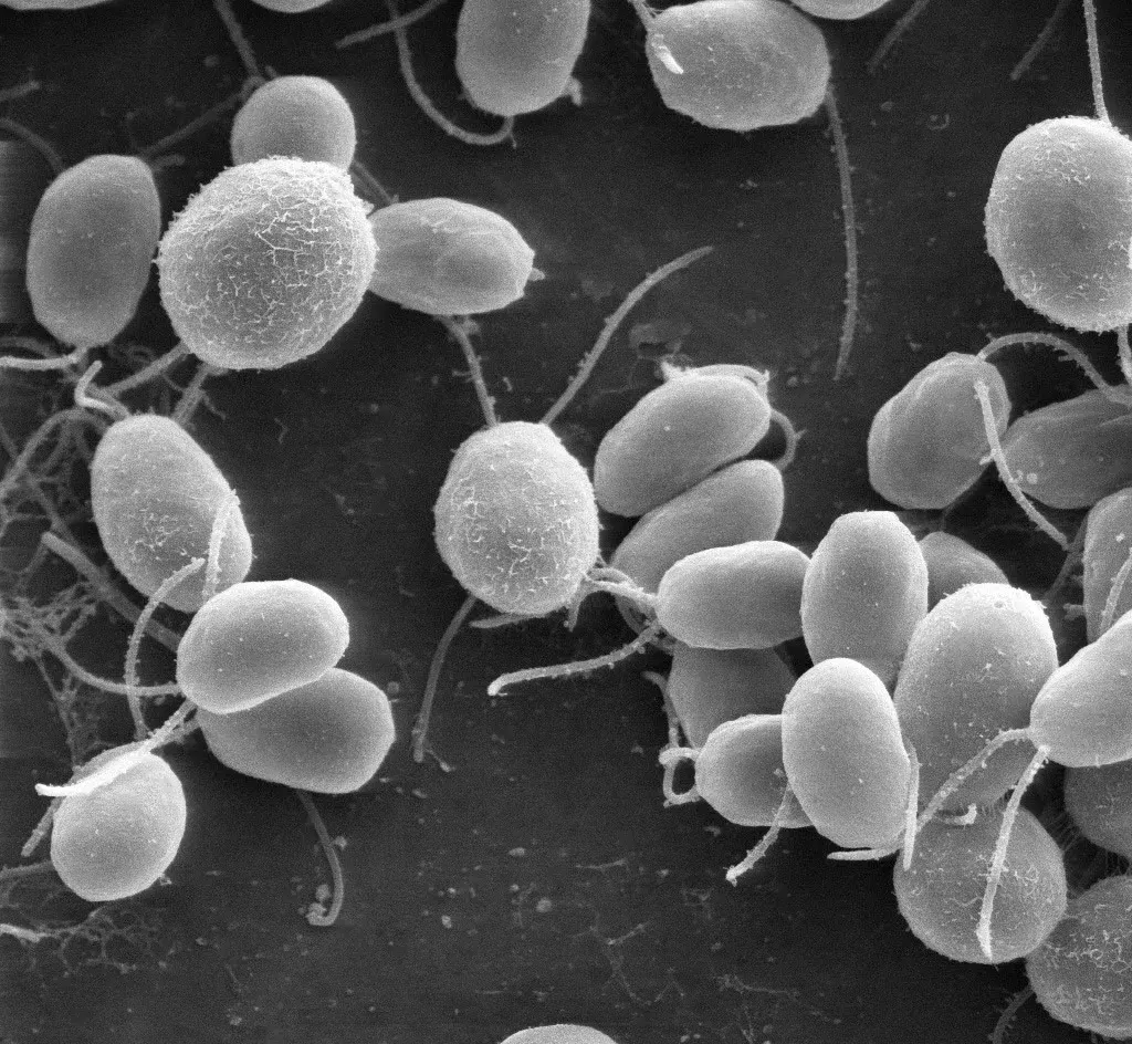
Figure: The single-celled alga Chlamydomonas reinhardtii under a scanning electron microscope, each cell with two flagella (2007) · Public domain, via Wikimedia Commons
This article follows the path that single protein has walked. From pond swimming to the human eye, it is a journey of just over twenty years.
2002, A Door Where Light Also Serves as the Handle
Cells are surrounded by thin membranes, and those membranes have doors built into them. An ion channel is a protein embedded in the thin membrane surrounding a cell, acting as a gate that opens and closes to let electrically charged particles pass through. It is a side door built into the membrane—when it opens, the electrical state inside and outside changes. Usually, these doors open in response to other signals. Some substance must come and attach, or the electrical state of the membrane must change first.
There are also proteins dedicated to handling light. Microbial rhodopsins are a family of light-receiving proteins found in single-celled organisms like algae and bacteria. Within this family, some pumps push protons across one side of the membrane in response to light, while other pumps push chloride (PMID 16042615).
Pumps and doors do different jobs. A pump pushes particles in one direction, thereby building up a difference between the inside and the outside. A door merely opens, and particles flow on their own according to the difference that has already been built up. Pushing requires effort, but opening the door does not require that kind of effort.
The path opened up in the data. Once the entire genome sequence of this green alga was read and the data made public, several research teams were able to point out candidate sequences that appeared to be light-receiving proteins within it (PMID 16042615). It was the exact moment when the question of observing swimming in a pond shifted to the question of scouring lists of sequences.
In 2002, the sequence of a new gene belonging to this family was reported from a green alga called Chlamydomonas reinhardtii (PMID 12089443). The core part of the protein pointed to by the sequence resembled a light-operated proton pump. However, when that gene was inserted into the cells of another living thing and exposed to light, it did not act like a pumping pump, but instead operated as a channel that selectively let only protons pass through (PMID 12089443). It had a body resembling a pump, but it did the work of a door.
At this point, a new name became necessary. Channelrhodopsin is an ion channel that opens by itself and allows ions to pass through when it receives light. It is a door where light also serves as the handle.
This first channelrhodopsin is made of 712 amino acids, and just about the front half of it was enough to operate as a light-responsive channel. The light that triggers the strongest response is around 500 nanometers, which is toward the green light spectrum (PMID 16042615). The first author of the 2002 paper is Georg Nagel, and the last author is Peter Hegemann.
The research that started out to explain the swimming direction of algae delivered something useful for all of biology right here. It is a door that opens and closes with just light.
2003: A Gateway That Lets Cations In
The following year, a second channel was reported in the same algae. It is channelrhodopsin-2. It consists of 737 amino acids, and the light to which it reacts most strongly is around 460 nanometers, which is on the bluer side than the first one (PMID 16042615).
The difference more important than color was what it allowed to pass through. The first channel let only protons pass, but the second channel widely let cations—charged particles—through. Monovalent and divalent cations passed together (PMID 14615590). When a single photon of light is absorbed, it opens immediately (PMID 14615590). It does not go through other signals in between.
The more decisive fact was where it worked. This channel worked in frog egg cells and in mammalian cells as well (PMID 14615590). If you put a single algal gene into an animal cell, that animal cell comes to react to light. The 2003 paper notes at the end that with this protein, simply shining light on cells—whether small or large—can change their electrical state (PMID 14615590).
There was something easily confusing about its shape. This structure crossing the membrane seven times is a characteristic of the receptor family that receives signals from outside the cell and transmits them inward, but its actual function was a channel that directly lets cations pass through (PMID 14615590). This meant it belonged to a family with a matching outward appearance, yet did a different job.
The limitations were also recorded together in the same report. If light is shone continuously, the channel desensitizes, and the current settles into a small state (PMID 14615590). Within the algae, this channel was produced mainly in environments where light was weak (PMID 14615590). The first author of the 2003 paper is also Georg Nagel, and Peter Hegemann is also listed as an author.
The molecule brought out to explain the life of algae changed its character at this point. Inserting a single gene and turning on the light—just these two things made it possible to tinker with the electricity inside someone else's cells.
What a Single Flash of Light Does Inside a Cell
With just the facts covered so far, we can follow a single scene. It is a scene where a cell bearing this channel is flashed with blue light for a very brief moment.
When a single light particle is absorbed by the channel, the channel opens immediately (PMID 14615590). When the door opens, positive ions flow—sodium, potassium, protons, and calcium stream in together (PMID 39594600). Because electrically charged particles enter, the electrical state across the membrane changes. A nerve cell is a fundamental cell of the brain and nerves that receives electrical signals and passes them on to the next cell. In that cell, this change in the electrical state acts as a trigger that fires a short electrical signal (PMID 39594600).
When the light is turned off, the channel closes. The speed at which it closes slows down depending on the amount of protons inside the cell (PMID 14615590). Looking at the links of the chain one by one, there are no missing steps in this scene. Light—opening—ion flow—change in electrical state—signal. This five-step path is how a single molecule from algae touches the electricity of an animal cell.
The same scene also shows the weakness of this method. It fits well for uses where the light is flashed in short bursts, but if left on continuously, the channel becomes desensitized and the current subsides (PMID 14615590). The recovery speed from this desensitized state becomes faster as there are more protons outside the cell and as the inside of the membrane is more negatively charged (PMID 14615590). A switch that is easy to handle and a light that stays on for a long time are different things, and this first-discovered channel is closer to the former.
2005, Turning on Cells in Milliseconds
Neurons do not send signals as continuous streams of intensity. They send short electrical signals by bursting them one by one, and each individual signal unfolds in units of 1/1000 of a second. Which neuron bursts a signal at which moment is precisely the language the brain speaks.
In 2005, a report emerged detailing the introduction of channelrhodopsin-2 from algae into mammalian neurons (PMID 16116447). The blueprint for this light-responsive protein was inserted into neurons, and a device that rapidly turns light on and off was attached. The result was twofold. It reliably controlled the timing of neuronal signal bursting at the millisecond scale, and the crossing of signals between neurons—both the side pushing the next cell and the side suppressing it—was controlled in tandem (PMID 16116447).
Optogenetics is a method that inserts the gene for a light-responsive protein into targeted cells, turning the activity of those cells on and off with light. The gene must enter first. Merely shining light makes nothing happen.
The other pillar of this method is "which cells it is put into." When introducing the gene, if it is tailored to operate only in a specific type of cell, the cells that react while the light is shone are also limited to that type. This is where the title of the 2005 report noted "genetically targeted" control (PMID 16116447). Even if the light spreads widely, the only cells that react are those that received the blueprint. The task of weaving together both at the molecular and optical levels which specific cells are allowed to react remains on the list of challenges today (PMID 41254285).
Why the unit of a millisecond matters becomes clear at this juncture. To figure out what a given population of cells does in the brain, one must be able to select and turn that exact population on and off at will. If the control is slow, one can only say "when this cell turned on, that event happened." When control becomes fast down to the scale of a single signal, it becomes possible to handle sequences. The first author of the 2005 paper was Edward Boyden, the final author was Karl Deisseroth, and Georg Nagel participated as a co-author.
The sequence in 2002, the channel in 2003, the neuron in 2005. As three reports spanning three years followed one another, a single molecule from pond algae became a switch for studying the brain. The path pointed to by the 2026 prize consists of these three steps. The original text for the award citation is "for their discoveries concerning light-gated ion channels and optogenetics," meaning discoveries concerning ion channels opened and closed by light and optogenetics (Nobel Committee, press release). According to the Nobel Committee's general background materials, this method acquired the name optogenetics in 2006, and was used in the brains of living mice the following year (Nobel Committee, popular background).
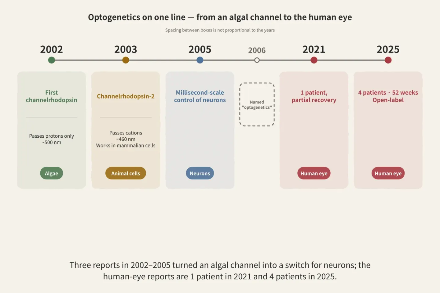
Figure: a diagram Paperis drew using only what the article says
Tools Multiply
The channelrhodopsins first discovered were pathways that let cations pass indiscriminately. Sodium, potassium, protons, and calcium flow in together, and the resulting change in the membrane's electrical state causes nerve cells to fire signals (PMID 39594600). The property of not being selective was convenient to use while at the same time crude. This is because unwanted particles move along together.
So, over the span of 20 years, this protein grew from a single entity into an entire family. Channelrhodopsins that selectively let only sodium through were found in nature and also built by human hands, and some of them hardly desensitized even when the light stayed on continuously (PMID 39594600). Variants with shifted light colors also emerged. Variants operating at longer wavelengths, meaning light closer to the red end, reduced the strain that light itself places on tissues; other variants achieved faster responses; and still others were tuned to operate under ordinary ambient brightness (PMID 40002842).
Why the indiscriminate property is crude becomes clear when you think about desired uses. The goal is to switch on only specific cells at the desired moment, but if multiple types of particles move together every time they are switched on, other conditions inside the cell are disrupted along with them. Pathways that pass only sodium can push the electrical state strongly while reducing that collateral movement (PMID 39594600).
Looking at light-responsive proteins as a whole, there are two branches. One comes from single-celled organisms and makes ions flow across the membrane when it receives light. The other is a protein originally found in the mammalian retina that starts a signal transduction process inside the cell upon receiving light (PMID 31982136). Over the past decade or so, the branch widely used in attempts to restore eye function has been the former one (PMID 31982136).
The latter branch is also under consideration as a candidate. Because of expectations that proteins originally found in the mammalian retina are more sensitive to light and better tolerated by the body, they have been discussed relatively recently as strong candidates for vision restoration (PMID 31982136). The method of receiving light to immediately flow ions and the method of receiving light to start signal transduction inside the cell have different balances of speed and sensitivity.
Thanks to the multiplication of tools, one condition has changed. With the advent of microbial proteins that are more sensitive to light and operate further toward the red spectrum, a path has opened up to stimulate cells at a brightness safely below human eye safety standards (PMID 25759964).
What Has Changed
Since the story of the tools has grown long, we must write down what came to be done with those tools. Optogenetics has transformed basic research on neural circuits, leading to various experimental insights into human brain function and its disorders — as a 2025 review paper summarizes (PMID 41254285).
What has changed becomes apparent when we look at the shape of the questions. When you cannot turn a specific group of cells on and off at will, the most observation can achieve is “when these cells are active, that behavior appears alongside them”. Events appearing together do not tell you which side is the cause. Once it becomes possible to selectively turn just that group on and off, the question changes to “does turning these cells on make that behavior appear?”. The same review notes that new understanding of how specific cell types contribute to cognition and behavior came from there, and that this understanding opened paths toward treatment (PMID 41254285).
There is a place to be careful here. A significant portion of these insights was obtained from non-human animals, not by directly putting light and genes into human brains. That is also why the same review divides the path toward treatment into two parts (PMID 41254285).
What the tools changed is the kind of questions you can ask. The place where those questions cross over into the human body must be viewed separately.
The Path Taken Through the Eyes — One Person, and Four People
The eye is the place where the method of controlling cells with light was first tested inside the human body. Three conditions align in the eye. The pathway to let light in is naturally open, the range over which to spread the carrier loaded with genes is small, and one can look in from the outside to check what has happened. Deep inside the body, simply sending the light becomes a separate homework assignment to solve first.
Retinitis pigmentosa is a genetic disease in which the light-receiving cells gradually die in the retina, the light-receiving membrane at the back of the eye. Grouped together, these hereditary retinal disorders affect about 1 in every 3,000 to 5,000 people, and are currently considered the most common cause of visual impairment registration in developed countries (PMID 30218651). At the stage where the light-receiving cells have died and vanished, a path of making other cells remaining in the retina respond to light in their place is being explored (PMID 30218651). A feature of this approach is that it can be applied regardless of which gene is broken (PMID 30218651).
Carriers take charge of sending genes into cells. A viral vector is a carrier made by taking a virus shell emptied of its disease-causing genes, loading it with the desired gene, and having it deliver it into the target cell. It is a delivery truck loaded with cargo instead of its original load.
The first reported result applied to humans is a case report of a single patient in 2021 (PMID 34031601). The gene for a protein that responds to reddish light was introduced via a viral vector into the eye of a patient who had lost vision due to retinitis pigmentosa, and special goggles that capture changes in light intensity and project them onto the retina as light pulses were worn together. With the treated eye and the goggles used together, this patient was able to recognize objects, point to where they were, count them, and pick them up with their hand (PMID 34031601). The same report also records that the signal went all the way to the brain. While the patient recognized objects, electrical activity related to the objects was recorded over the brain region responsible for vision (PMID 34031601). This means the light that entered the eye did not stop at the eye.
The reason goggles are needed lies on the protein side. The proteins used today do not create usable vision with ordinary indoor brightness alone, and thus a device that gathers light and increases its intensity was used in tandem. Making them work in ordinary brightness without an external device is cited as a line this field has not yet crossed (PMID 40002842).
According to the same report, this patient could not see objects before the injection whether wearing goggles or not, and could not see without goggles even after the injection (PMID 34031601). This is a case report of a single patient, and what was recovered is partial function.
In 2025, a report on four patients came out (PMID 40121528). The gene for a new protein created by modifying and combining three kinds of non-human biological proteins was loaded into a viral vector and given as a single injection targeting a specific cell layer inside the retina. The subjects were four retinitis pigmentosa patients with a specific genetic mutation, and improvements in vision, shape discrimination, and movement were reported up to 52 weeks, with no obvious safety issues reported (PMID 40121528). The authors conclude that this report is the first report of standalone gene therapy to restore vision regardless of specific genetic mutations (PMID 40121528). This trial is an open-label single-arm study without a comparison group, where both participants and researchers know what was received. The denominator of four people and the duration of 52 weeks are the scope of this result.
This report also records the verification process. Whether the introduced gene was actually expressed in the retina was checked by observing it through non-invasive imaging using a co-delivered fluorescent reporter protein (PMID 40121528). The same paper notes that this protein is likely one of the most artificial non-mammalian proteins ever expressed in humans, while adding that there were nevertheless no clear safety issues (PMID 40121528).
Aside from these two reports, trials are ongoing. Two clinical trials aiming to restore light sensation in retinitis pigmentosa patients using microorganism-derived proteins were underway as of 2020 (PMID 31982136), and a review from earlier in 2019 also notes that one phase 1/2 trial is underway (PMID 30218651). Trials before results come out are neither expectations nor conclusions.
The limitations of the proteins currently used are also recorded in the same report. The response is slow, they are already partially turned on at ordinary surrounding brightness, they cannot respond by distinguishing colors of light, and the targeted retinal circuit is crude in terms of resolution (PMID 40121528). Which combination among the proteins, carriers, and surgical methods to use for the eye has not yet been decided either (PMID 30218651).
Who it will be used for is also open. Retinitis pigmentosa and age-related retinal diseases resemble each other in that light-receiving cells die, but the number of patients is vastly larger on the age-related disease side (PMID 39640234). Even so, research resources have been poured more heavily into retinitis pigmentosa, where disease models are easier to handle, and many blanks remain regarding how results obtained on one side will translate to patients on the other (PMID 39640234). The problem where the number of patients and the easy-to-study models point in opposite directions remains here.
To sum up what happened in the eye in one line: a report has emerged that partially restored human vision using an avian protein, and the denominator of that report is still in the single digits.
What We Still Do Not Know
The path toward human treatments branches in two directions. One is placing light and genes directly into the human body, and the other is using causal knowledge gained from animals in other therapeutic means. A 2025 review summarizes that a significant portion of the paths opened so far belongs to the latter — knowledge uncovered through optogenetics about which cells handle which functions has flowed into the design of non-optogenetic therapies (PMID 41254285). The same review notes that direct application cases emerging from the eye have revealed the need for careful ethical review alongside therapeutic possibilities, and lists choosing which diseases to target, how to aim at specific cells only, and how to pass safety and regulation as remaining challenges (PMID 41254285).
The blanks on the tool side are relatively clear. More efficient and safer carriers, proteins shifted further toward the red spectrum, materials that deliver light close to infrared deep into tissue, and implantable devices that send light deep inside the body without wires are cited as necessary items on the list (PMID 35027688). In the eye, the task of creating usable vision with ordinary indoor brightness alone without external devices remains a challenge (PMID 40002842).
Looking at the nature of the list reveals where the remaining problems are concentrated. Rather than the light-responsive proteins themselves, the challenges are clustered around safely getting them into the desired cells and sending light deep into the body. Conversely, that is why the eye opened first — in other locations, both delivery and illumination must be solved anew.
For uses outside the eye, some are still at the proposal stage. A delivery method of placing a sodium-selective channel into red blood cells and shining light to release drugs was proposed in a review (PMID 39594600). It is a proposal, not a result used in humans.
The ear is discussed as the location after the eye. The cochlear implant is cited as the most successful neural prosthetic device, and the majority of 1 million patients who would otherwise not have been able to hear sounds have come to understand speech (PMID 38594945). However, when stimulating the auditory nerve with electricity, the current spreads widely, so the pitch information that can be transmitted gets blocked there. The same review notes that while the cochlear implant is cited as the most successful device, a large unmet medical need remains in raising the quality of hearing (PMID 38594945). Expectations arise that light can be focused into a narrower range than current and thus stimulate with greater precision, but the review evaluates that many challenges remain before it can be used in humans (PMID 38594945).
Gathering the human-subject reports written so far into one place, the numbers are small. One patient in 2021 and four patients in 2025, both retinitis pigmentosa, both the eye. Both are reports tracking only the recipients without a comparison group, and the 2021 side had to wear special goggles together (PMID 34031601, PMID 40121528). The position this method occupies as a therapy used in humans is told by this denominator.
Although the numbers reported in human treatments are still small, as a research tool this method has already changed neural circuit research and opened several windows for viewing the function and diseases of the human brain (PMID 41254285). The algal protein that once swam in a pond is currently used most widely in laboratories. Its use in hospitals follows far behind that.
Where to Read Further
Why turning things on and off with light is so useful becomes sharp and clear when you know how nerve cells send and receive signals. "How Do Nerve Cells Send Signals? — A Burning Fuse, a Gap Crossed by Ferry" is that story. The point that putting in a single gene makes that cell produce a new protein continues in "From Gene to Protein — A Copy Read Three Letters at a Time, and the Pages Each Cell Opens". How far the work of shaping and making proteins by human hands has come these days can be seen in "From Prediction to Design — After AlphaFold, the Science of Drawing Proteins and Building Them". What happens in the body when nerve cells misfire all at once is covered by "What Is Epilepsy? — Windows Lit One by One, Lights That Flash All at Once".
Awards in other categories that same year continue in The 2026 Nobel Prize in Physics — Messengers from the Cosmos, Caught in a Cubic Kilometer of Ice, The 2026 Nobel Prize in Chemistry — Chemistry That Makes One Hand on Its Own, and Did Life Take That Path?, The 2026 Nobel Prize in Economics — From One Household's Arithmetic to a Nation's Business Cycle, The 2026 Nobel Prize in Literature — Anne Carson, New Forms from a Dialogue with Ancient Poetry, and The 2026 Nobel Peace Prize — Crimes Named in Court: Navi Pillay and the International Criminal Tribunal for Rwanda.
Sources
- "2026 Nobel Prize in Physiology or Medicine Press Release" (2026) — https://www.nobelprize.org/prizes/medicine/2026/ (Karolinska Institute Nobel Assembly: https://www.nobelprizemedicine.org/the-nobel-prize-in-physiology-or-medicine-2026/) — Laureates, affiliations, reasons for the prize, and the roles of the three laureates
- "2026 Nobel Prize in Physiology or Medicine Popular Background" (2026) — https://www.nobelprizemedicine.org/wp-content/uploads/2026/10/Nobel_Prize_ENG_pop.pdf — Naming of "optogenetics" in 2006, application to mouse brains in 2007
- "2026 Nobel Prize in Physiology or Medicine Advanced Background" (2026) — https://www.nobelprizemedicine.org/wp-content/uploads/2026/10/Nobel_Prize_ENG_adv.pdf — Scientific background of the discovery
Source Papers
- "Channelrhodopsin-1: a light-gated proton channel in green algae" (2002) — PMID 12089443 — Green algae's phototactic and photophobic responses are mediated by microbial rhodopsins, and when sequences found in Chlamydomonas were put into other cells, a light-operated channel that selectively lets through only protons appeared.
- "Channelrhodopsins: directly light-gated cation channels" (2005) — PMID 16042615 — The first channelrhodopsin has 712 amino acids and a structure spanning the membrane seven times, with a peak around 500 nanometers and proton selectivity. Channelrhodopsin-2 has 737 amino acids and a peak around 460 nanometers on the blue side, passing monovalent and divalent cations.
- "Channelrhodopsin-2, a directly light-gated cation-selective membrane channel" (2003) — PMID 14615590 — A directly light-gated cation-selective membrane channel that works in both frog oocytes and mammalian cells, opening immediately after photon absorption, desensitizing in continuous light, and able to depolarize cells with light alone.
- "Millisecond-timescale, genetically targeted optical control of neural activity" (2005) — PMID 16116447 — Used the algal protein channelrhodopsin-2 and a high-speed optical switch to control the firing timing of mammalian neurons on a millisecond scale and regulate excitatory and inhibitory synaptic transmission as well.
- "Sodium-Selective Channelrhodopsins" (2024) — PMID 39594600 — Early channelrhodopsins were cation channels indiscriminately passing sodium, potassium, protons, and calcium, after which sodium-selective variants were discovered and engineered, some showing almost no desensitization in continuous light.
- "Evolution of Light-Sensitive Proteins in Optogenetic Approaches for Vision Restoration: A Comprehensive Review" (2025) — PMID 40002842 — A summary of the evolution of variants from ReaChR and ChrimsonR shifted toward the red side, Chronos with fast kinetics, to MCO1 operating in ambient light, and the remaining task of achieving functional vision at ordinary indoor brightness without external devices.
- "Opsins for vision restoration" (2020) — PMID 31982136 — Opsins that regulate membrane potential fall into two branches: microbial opsins derived from single-celled organisms and animal opsins from the mammalian retina, with microbial opsins being the ones widely used for vision restoration over the past decade or so.
- "Optogenetics" (2015) — PMID 25759964 — Microbial opsins with improved light sensitivity and action spectra shifted toward the red side have made it possible to stimulate at brightnesses sufficiently lower than the human eye's safety limits.
- "Optogenetic approaches to vision restoration" (2019) — PMID 30218651 — Inherited retinal diseases affect 1 in 3,000 to 5,000 people and are the most common cause of registered visual impairment in developed countries, forming a gene-agnostic strategy that makes secondary and tertiary retinal neurons respond to light, though optimal molecules, vectors, and surgical methods are not yet settled.
- "Partial recovery of visual function in a blind patient after optogenetic therapy" (2021) — PMID 34031601 — A single patient was injected in the eye with a vector carrying ChrimsonR and used special goggles together to recognize, count, and pick up objects, which they could not see before the injection and without the goggles.
- "A synthetic opsin restores vision in patients with severe retinal degeneration" (2025) — PMID 40121528 — In an open-label single-arm study where a synthetic opsin engineered and combined from three non-mammalian proteins was delivered to retinal bipolar cells via AAV2, four patients showed improvements in vision, shape distinction, and movement up to 52 weeks, along with notes on the limitations of current opsins (slow kinetics, partial activity in ambient light, lack of color response, low resolution).
- "Bridging the gap of vision restoration" (2024) — PMID 39640234 — Retinitis pigmentosa and age-related retinal degeneration resemble each other in the loss of photoreceptor cells, but the latter has a larger number of patients, whereas more resources are poured into the former because its models are easier to handle, leaving many blanks in the transition between the two groups.
- "Roadmap for direct and indirect translation of optogenetics into discoveries and therapies for humans" (2025) — PMID 41254285 — Optogenetics has transformed basic neural circuit research, and much of the path toward therapy relies on utilizing causal knowledge rather than direct application, with direct applications to the human central nervous system revealing both therapeutic potential and the need for ethical review.
- "Towards translational optogenetics" (2023) — PMID 35027688 — The toolbox consists of light-responsive proteins, delivery vectors, and light-delivery devices, with safer vectors, proteins shifted further toward the red side, nanomaterials delivering near-infrared light deeply, and wireless implantable light-delivery devices listed on the wish list.
- "Toward Optogenetic Hearing Restoration" (2024) — PMID 38594945 — Cochlear implants are the most successful neural prosthetic devices that enable speech understanding for the vast majority of patients on a scale of 1 million, but current spread is the bottleneck for transmitting frequency information, and light can be narrowly focused spatially so it is proposed as an alternative, though challenges remain until clinical application.