Paperis Articles
Girl with a Pearl Earring — Light Built in Layers, a Name Not Asked
In 2018 The Girl in the Spotlight project examined Vermeer's painting of around 1665 almost without touching it. We follow what pigment maps, the preparatory stages beneath the surface, ultramarine and lead white, the darkened background and the surface cracks answered about how it was painted — and the question the project did not ask.
Your field's papers, as audio
Pick your fields and journals — Paperis summarizes the key papers and turns them into audio. Listen on your commute.
Start Paperis free →Girl with a Pearl Earring — Light Built in Layers, a Name Not Asked
Around 1665, now The Hague
The Dutch painter Johannes Vermeer was born in 1632 and passed away in 1675 (DOI: 10.1186/s40494-020-00364-5). He worked in the Dutch city of Delft. "Girl with a Pearl Earring" is a painting he created around 1665. Against a dark background, a young woman wrapped in a blue and yellow headscarf turns her head over her shoulder to look this way. Her lips are slightly parted, and a large pearl earring hangs from her ear. The canvas size is 39×44.5 centimeters (DOI: 10.2312/gch.20181336).
This painting is now in the Mauritshuis museum in The Hague, Netherlands. An overview paper of the museum-led investigation introduces this painting as the most beloved among the museum's collection (DOI: 10.1186/s40494-019-0307-5).
It is not known who the woman in the painting is. This painting is often classified not as a portrait of a specific individual, but as a tronie, that is, a character study.
Before 2018, the last time this painting was examined was during a 1994 restoration. At that time, conservation specialists and scientists used visible light, ultraviolet, and infrared photography, X-ray photography, and stereomicroscopes, and cut and looked at the cross-section of tiny paint flakes removed from the painting (DOI: 10.1186/s40494-019-0307-5). In 2018, an investigation titled "Girl in the Spotlight" was held. It was an investigation led by the Mauritshuis, within the framework of NICAS, a Dutch collaborating agency for conservation, art, and science, together with internationally recognized specialists and scientists from other institutions. The painting was examined inside the museum right in front of visitors (DOI: 10.1186/s40494-019-0307-5).
This article follows what the 2018 investigation found in the painting. What the investigation answered is "how this painting was made and how it has changed," while "who was painted and why" are questions the investigation did not ask.
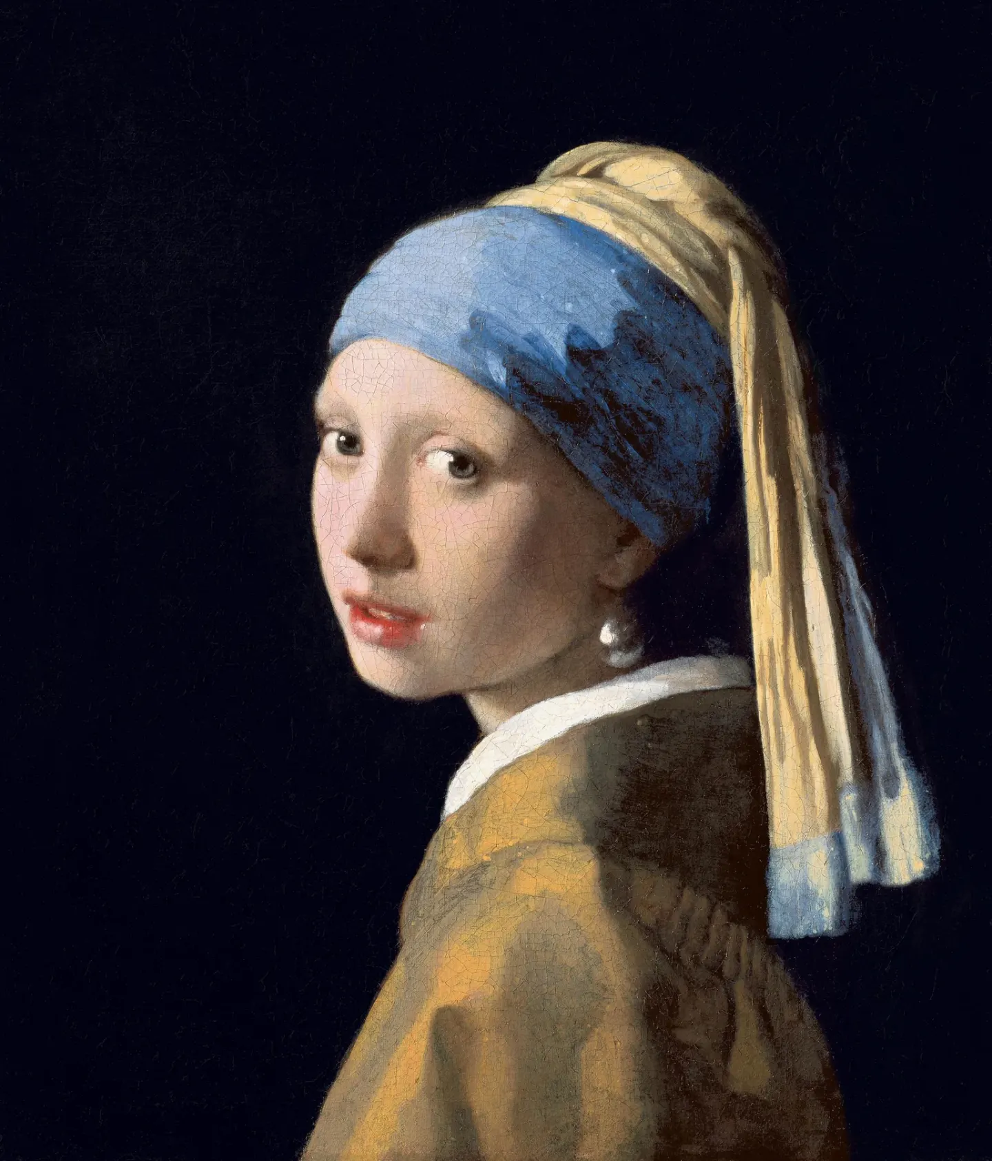
Johannes Vermeer, Girl with a Pearl Earring (c. 1665) · Mauritshuis, The Hague · public domain · Wikimedia Commons
How to Measure Without Touching the Painting
The questions were five. What can be found in the layers beneath the surface. What steps Vermeer took to paint this picture. What materials were used and where those materials came from. By what technique subtle light effects were created. How it looked at first and how it changed afterwards. The 2018 investigation began by tackling these five (DOI: 10.1186/s40494-020-0359-6).
To answer these questions, the research team overlapped several methods that barely touched the painting. Infrared photography, spectral imaging that captures reflection and fluorescence by light wavelength, 3D scans, 3D digital microscopes, and X-ray scanning were included, and a paint chip detached in 1994 was also reanalyzed with new equipment (DOI: 10.1186/s40494-019-0307-5). The 3D digital microscope magnified up to 140 times to take photographs where one pixel is 1.1 micrometers, a fraction of the thickness of a human hair (DOI: 10.1186/s40494-020-0359-6).
Among them, two appear frequently in this text. Macro XRF is a method that scans across the painting with X-rays, receives the light returning differently for each element, and maps out which element is in which spot. Spots high in lead and spots high in calcium appear like a color map across the entire painting. Optical coherence tomography is a method that shoots light and compares the returning light to measure the height of the painting's surface and the thickness of the transparent layer just below it without cutting. In this investigation, these scans were automatically stitched together to photograph an entire painting in micrometer units, and that data revealed brushstrokes, surface cracks, places where paint flaked off, and past restoration marks (DOI: 10.1364/oe.390703).
The 2018 investigation imaged and identified the materials on the painting's surface and underneath it, making it possible to overlap and compare the results of multiple instruments at the exact same spots (DOI: 10.1186/s40494-019-0307-5). Therefore, many of the results appearing in this text are maps plotting "what is where."
Beneath the Surface: The Gray Ground, Black Lines, and Underpaint
The canvas is painted with a warm, light gray ground, and this ground was probably not applied by Vermeer himself (DOI: 10.1186/s40494-019-0308-4).
An underpaint is a layer of paint laid down first in that spot before the final colors go on top. The tinted gray ground served as a mid-tone background, and Vermeer began laying in the light and dark areas of the composition over it, while also drawing a few thin contour lines around the figures (DOI: 10.1186/s40494-019-0308-4). The underpaint in this painting varies in color and thickness from place to place, with the upper paint layers applied on top of it (DOI: 10.1186/s40494-019-0308-4).
Most of this preparation stage is hidden beneath the surface, so the research team peeked inside using infrared light. Infrared light penetrates the upper paint layers to some extent to reveal the black lines underneath. By combining microscope images with infrared photographs taken at four wavelength bands from 900 to 2,500 nanometers at 0.05-millimeter intervals, the preparation stage was found to consist of three parts: the gray ground, thin black outlines, and the underpaint (DOI: 10.1186/s40494-019-0308-4). The paper's abstract simply lists these three without stating whether the outlines or the underpaint came first. In spots where the underpaint was left exposed—such as around the figures—and where the upper layers are thin, this underpaint is still visible under a microscope (DOI: 10.1186/s40494-019-0308-4).
Signs of changing one's mind midway also emerged. There were several places where changes were made after painting, and one of them is the position of the ear (DOI: 10.1186/s40494-019-0308-4). The research team also used a computer to count the canvas threads captured in X-ray photographs to measure thread spacing and angles.
The research team's conclusion is that the materials beneath the surface played a major role in determining the depth of the space within the painting, the direction from which light falls, and the final colors in various parts of the painting (DOI: 10.1186/s40494-019-0308-4). In many areas of this painting, the colors we see are shaped in large part not just by the topmost layer, but also by the materials underneath it.
Blue Headscarf and Ultramarine
One striking color in this painting is the blue of the headscarf. Ultramarine is a blue pigment made by grinding a blue stone called lapis lazuli. Pigments refer to powders that give paint its color. Vermeer was known for his bright blue and frequently used expensive natural ultramarine (DOI: 10.1186/s40494-020-00364-5).
The ultramarine in the headscarf of this painting was of high quality. When looking at a cross-section of a paint sample with an electron microscope and infrared spectroscopy, there were many mineral grains producing a bright blue color (DOI: 10.1186/s40494-020-00364-5). An analysis looking at the state of sulfur atoms using a synchrotron particle accelerator suggested that this ultramarine was made, at least in part, from lapis lazuli heated in a fire (DOI: 10.1186/s40494-020-00364-5). The word chosen by the authors is "suggests."
A map of the entire headscarf also emerged. A white pigment appears alongside it here. Lead white is a white pigment made from lead. The blue parts of the headscarf were painted by mixing natural ultramarine with varying amounts of lead white (DOI: 10.1186/s40494-019-0348-9). The yellow parts were painted with yellow earth pigment and lead white, and the edge of the headscarf contained additional ultramarine and a yellow made of lead and tin (DOI: 10.1186/s40494-019-0348-9). This investigation confirmed previous findings that Vermeer used only a handful of pigments while mapping out where and how each pigment was used (DOI: 10.1186/s40494-019-0348-9).
However, the shadow side of the headscarf is noticeably mottled. The research team considers this to be the result of degraded paint, likely related to the chalk that Vermeer mixed heavily into the ultramarine paint in that spot (DOI: 10.1186/s40494-020-00364-5). Chalk is a white calcareous powder. A question arises from this. It is whether Vermeer intentionally added chalk to adjust the workability or transparency of the paint, or whether that chalk was a base powder used to anchor a yellow colorant that has now faded (DOI: 10.1186/s40494-020-00364-5). The paper leaves this question open.
A glaze is a thin, transparent layer of paint applied on top so that the color underneath shows through. The research team made reproduction paints containing chalk or yellow colorant and compared their properties. What is presented by combining the analysis and the reproduction paint experiments is a hypothesis. It is that the blue of the headscarf originally fell into three branches: the left side receiving light was an opaque bright blue, the middle was a slightly more vivid opaque blue, and the shadow side was a color made by alternating deep, dark cyan glaze with cyan brushstrokes, and that the color on the shadow side has now mostly been damaged due to paint degradation (DOI: 10.1186/s40494-020-00364-5).
Therefore, the headscarf shadow we see now may not be the same as the shadow when it was first painted. However, the original appearance remains a hypothesis derived from analysis and reproduction paint experiments.
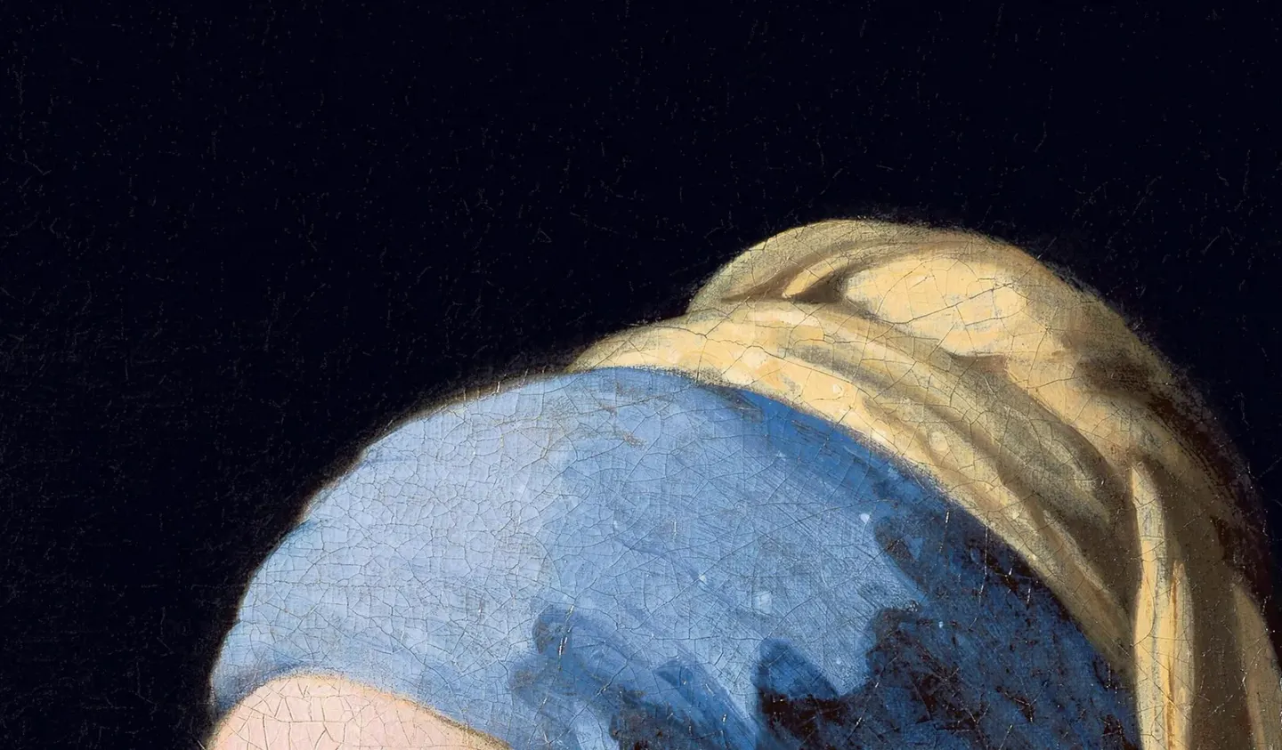
Detail: the headscarf · Johannes Vermeer, Girl with a Pearl Earring (c. 1665) · Mauritshuis, The Hague · public domain · Wikimedia Commons
Face: From Light and Dark Masses to Smooth Flesh
Art historians have pointed to the rendering of this girl's soft flesh, and that rendering is a main reason for dating the painting around 1665 (DOI: 10.1186/s40494-019-0344-0). This investigation peered into how that flesh was made.
The skin was painted with earth pigments, lead white, vermilion (a red mineral pigment), and a touch of red organic dye (DOI: 10.1186/s40494-019-0348-9). The fluorescence of the red organic dye was strongest on the lips, where vermilion also appeared (DOI: 10.1186/s40494-019-0348-9). The pearl earring was painted with a thinly rubbed layer of lead white and a lead white highlight (DOI: 10.1186/s40494-019-0348-9).
The order in which the face was painted also emerged. Vermeer first clearly divided the light and dark areas to build the face, and then smoothly blended the final layers on top so that the boundaries are barely visible (DOI: 10.1186/s40494-019-0344-0). The soft contour around the face was created by leaving a "gap" between the background and the flesh (DOI: 10.1186/s40494-019-0344-0). And details not visible to the naked eye, such as eyelashes, were revealed in the imaging (DOI: 10.1186/s40494-019-0344-0).
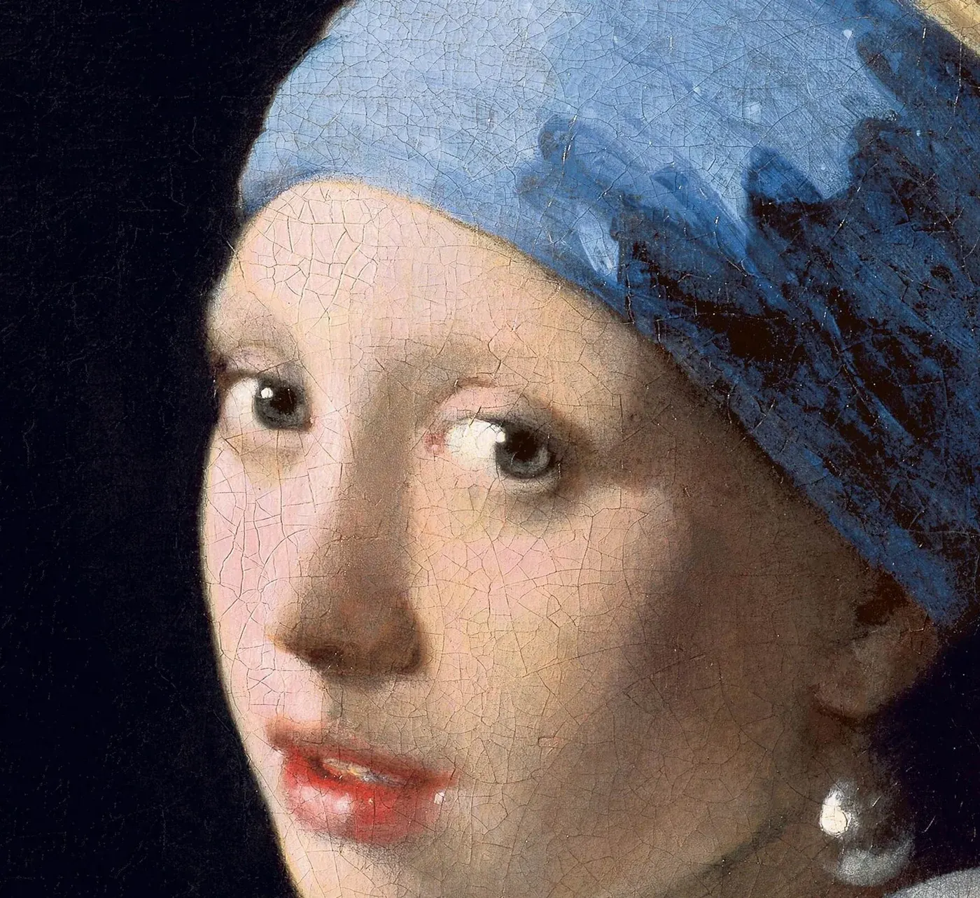
Detail: the face and pearl · Johannes Vermeer, Girl with a Pearl Earring (c. 1665) · Mauritshuis, The Hague · public domain · Wikimedia Commons
There were also differences in the white. The skin paint used lead white with differing ratios of crystals and particle sizes, and the authors suggest that Vermeer deliberately selected lead white of different qualities or grades (DOI: 10.1186/s40494-019-0344-0). When lead isotopes were analyzed, the lead used to make the different lead whites in this skin paint all came from a single place, the Peak District region in Derbyshire, England (DOI: 10.1186/s40494-019-0344-0). This is the result that lead whites made from lead of a single source were used in the skin paint with different grades.
Separately, another study that took X-ray images identifying materials by their crystal structure over a wide range on the painting and at a microscopic scale found four subtypes of lead white, and the authors read this to mean that Vermeer was very selective in using lead white (DOI: 10.1126/sciadv.aax1975). Whether these four are the same as the lead whites in the skin paint is not in the paper abstract.
Changes were also taking place within the facial paint. Cross-section analysis revealed newly formed lead compounds within the paint—namely, lead soaps and a mineral called palmierite—which were found alongside the red organic dye (DOI: 10.1186/s40494-019-0344-0).
Green Glow and Folded Cloth on a Dark Background
The background of this painting was interpreted as flat darkness until recently (DOI: 10.1186/s40494-019-0311-9). A 2018 examination of a cross-section of a paint fragment from the background showed that the darkness was two layers deep. Below is an undercoat mainly of charcoal black, and above is a glaze containing two organic colorants, blue indigo and yellow weld, with a copper-based drier (DOI: 10.1186/s40494-019-0311-9).
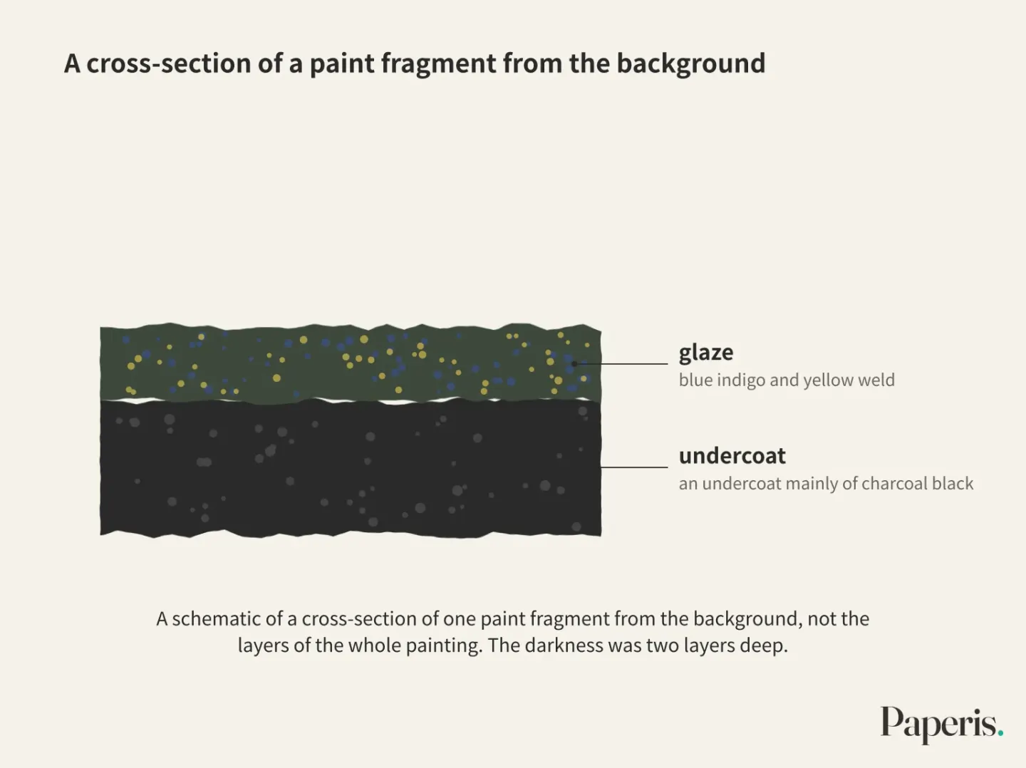
As that glaze degraded, the shapes in the background became hard to recognize with the naked eye (DOI: 10.1186/s40494-019-0311-9). Images capturing reflection by wavelength of light revealed the shapes of colors even though background reflectance was very low at 1 to 3%. A signal for the presence of indigo appeared, and while reflections of green wavelengths were not seen in most spots, the research team interprets this as an indication that the yellow weld had degraded (DOI: 10.1186/s40494-019-0348-9). However, green light emerged from a few small spots in the background. Those spots overlapped with areas that had been obscured by overpainting during past restorations, and thus appear to have been better preserved (DOI: 10.1186/s40494-019-0348-9). This green light is the result of a few small spots, and the paper's abstract does not state figures for how green the entire background originally was.
Images taken with overlapping exposures showed things that were hard to see with the naked eye. They are Vermeer's signature and traces of a folded cloth on the right side of the painting. The research team notes that this cloth "might be a curtain" (DOI: 10.1186/s40494-019-0311-9).
The painting technique was also revealed. Vermeer applied the black undercoat vigorously in overlapping brushstrokes of uneven thickness, and when he applied the glaze over it, the surface became smooth, flat, and even (DOI: 10.1186/s40494-019-0311-9). The research team believes this background creates an effect that contrasts with the girl, pushing her forward toward the viewer (DOI: 10.1186/s40494-019-0311-9).
The optical coherence tomography team reached the same conclusion. By adding optical properties measured by recreating Vermeer's paints in a historical manner to the measured surface and layer thicknesses, they created an on-screen painting where lighting, viewing angle, and the presence or absence of glaze could be changed. Based on that visualization, the authors conclude that the structure of the glaze layer and its optical properties were essential for Vermeer in creating a very strong contrast between the figure and the background (DOI: 10.1364/oe.390703). This is a conclusion that relies on measurements and visualizations made with recreated paints.
From Background to Cheek, Following a Single Line
Here, we gather the facts established so far into one spot on the painting. It is the edge of the girl's face, where the dark background meets the cheek. However, since the facts below come from different spots and methods, this is not a stratigraphic layer measured directly at this single boundary point.
In the cross-section of the paint fragment on the background side, an undercoat of charcoal black and an indigo and weld glaze above it were identified (DOI: 10.1186/s40494-019-0311-9). Whether this cross-section was lifted right from this boundary spot is not mentioned in the paper abstract. Across most spots of the background, signals that the yellow weld inside the glaze had degraded appeared (DOI: 10.1186/s40494-019-0348-9).
There is a gap at the boundary where the background and the skin meet (DOI: 10.1186/s40494-019-0344-0). There is also an observation that the undercoat is exposed like an outline around the figure (DOI: 10.1186/s40494-019-0308-4). The abstracts of the two papers merely describe this boundary as a "gap" and an "exposed undercoat outline," respectively, without stating whether the two observations are the same phenomenon at the same spot.
Cross the boundary, and you reach the skin. On top of a face built first with light masses and dark masses, the final layers are smoothly blended (DOI: 10.1186/s40494-019-0344-0), and lead whites with different crystal proportions and grain sizes were used in that skin paint (DOI: 10.1186/s40494-019-0344-0).
Gathered this way, different hands and times exist together in this painting. The gray ground of the canvas was probably applied by a hand other than Vermeer's, the flesh mixed with the undercoat is Vermeer's hand, and the degraded glaze of the background is a change that happened after painting.
The Chemistry of Surfaces, A Map Drawn by Cracks
New compounds that were not there at first have formed on the surface of the painting. When X-ray scanning, which identifies materials by their crystal structure, was run in a reflection mode more sensitive to the surface, the compounds close to the surface became clearly visible. Along with the original pigments, four newly formed compounds appeared: gypsum, the lead sulfate mineral anglesite, palmierite, and the calcium oxalate mineral weddellite (DOI: 10.1186/s40494-019-0309-3). The formation of gypsum was linked to the chalk contained in the upper glaze layer, and the formation of palmierite and weddellite was linked to the locations of the organic colorants and the base powder used to adhere those colorants (DOI: 10.1186/s40494-019-0309-3).
This map is also used to select where to detach paint fragments for further examination. When paint fragments detached from areas rich in palmierite were re-analyzed using synchrotron radiation, this compound was found at the boundary where the upper paint layer and the ground meet, and anglesite was also present in the ground layer (DOI: 10.1186/s40494-019-0309-3). The authors consider this scanning to be of great utility for evaluating the chemical condition of the painting (DOI: 10.1186/s40494-019-0309-3). Figures stating how fast these changes occur or how dangerous the painting is are not in the paper abstracts.
The shape of the surface also served as a map. 17th-century canvas paintings typically have a structure where varnish and translucent paint layers are superimposed on the canvas, and when cracks form within those layers, they appear in the surface relief (DOI: 10.1186/s40494-019-0331-5). The research team compared three 3D scanning methods. 3D digital microscopy and optical coherence tomography were the most accurate, but their field of view was narrow, making them too slow to capture an entire large painting (DOI: 10.1186/s40494-019-0331-5). The 3D data revealed aging manifested as moat-like depressions around thick impasto strokes, traces of past consolidation treatments for lifting paint flakes, and crack patterns. The authors view this data as a starting point for recording and monitoring how the surface changes in the future (DOI: 10.1186/s40494-019-0331-5).
The shape of those cracks was different from expectations. From a scanner that swept this painting at 7-micrometer intervals with a depth accuracy of 34 micrometers, cracks appeared more often as raised ridge shapes rather than inward-gouged valleys (DOI: 10.2312/gch.20181336).
There is also research that has read this crack network through formulas. One study, which applied a theoretical model of crack formation in dry films to images of this painting, estimated the thickness ratio of the left and right paint layers to be about 1.34 and the stress ratio to be about 1.102 in relation to the bright lighting effect on the girl's face (DOI: 10.1039/c6sm02849g). Another study calculated the disorder of the directions in which the cracks spread, reporting that the crack structure on the left side of the face is less ordered than the right, and suggests that if the experiment is expanded and applied to old paintings, that disorder has not yet reached saturation (DOI: 10.3390/e20100772). Both results are not direct measurements of the painting, but estimates obtained by overlaying theoretical models and sample experiments onto crack images.
What the results of this section tell us are the locations where changes occurred and their types. The speed of change and the magnitude of risk are not in these paper abstracts, and the surface map from 2018 serves as a baseline against which future investigations can be compared.
Questions Answered, Questions Left Unanswered
Layers, steps, materials and their origins, techniques, and original appearance versus changes. These are the five questions posed during the 2018 investigation (DOI: 10.1186/s40494-020-0359-6). The findings followed by this article answer those five one by one. There were three preparation steps—ground, outline, and underpaint—the ear position was corrected, high-grade ultramarine was used, different grades of lead white were used for the skin, the lead in the skin paint's lead white came from a single region in England, the face was blended from blocky shapes into smooth flesh, and the background and headscarf shadows have deteriorated.
Among the five questions, "Who is the woman in the painting?" is not included. Measurements answered "how it was painted" but do not move on to "why it was painted" or "who was painted." That the background pushes the girl forward and that glazes were essential for contrast are also interpretations by researchers standing on top of the measurements.
Even among the answers, certainties and hypotheses are mixed. Where the pigments are located was confirmed through maps, and the three preparation steps and background layers through infrared images, microscope photographs, and paint fragment cross-sections. The picture showing that the headscarf shadow was originally a deep teal glaze is a hypothesis derived from analysis and reconstructed paint experiments (DOI: 10.1186/s40494-020-00364-5), and whether chalk was intentionally added is an open question. The painting's creation date around 1665 is also an art-historical judgment based on this flesh representation (DOI: 10.1186/s40494-019-0344-0).
This painting can now be measured again in the future. The 2018 surface map serves as a baseline, allowing the next investigation to compare how the surface has changed in the meantime (DOI: 10.1186/s40494-019-0331-5).
Source Papers
- "From 'Vermeer Illuminated' to 'The Girl in the Spotlight': approaches and methodologies for the scientific (re-)examination of Vermeer's Girl with a Pearl Earring" (2019) — DOI: 10.1186/s40494-019-0307-5 — Overview of methods from the 1994 conservation survey and the 2018 in-front-of-visitors examination. One of the museum collection's most beloved paintings. Overlaying and comparing multiple imaging results.
- "The Girl in the Spotlight: Vermeer at work, his materials and techniques in Girl with a Pearl Earring" (2020) — DOI: 10.1186/s40494-020-0359-6 — Special issue synthesis. Five research questions, 140x 3D digital microscope (1.1 µm/pixel).
- "Revealing the painterly technique beneath the surface of Vermeer's Girl with a Pearl Earring using macro- and microscale imaging" (2019) — DOI: 10.1186/s40494-019-0308-4 — Light gray ground layer (likely not applied by Vermeer himself), black outlines, and three preparation stages of underpainting. Revision of the ear's position. Underlying materials play a major role in defining space, light, and final colors.
- "Out of the blue: Vermeer's use of ultramarine in Girl with a Pearl Earring" (2020) — DOI: 10.1186/s40494-020-00364-5 — High-quality ultramarine, some suggested to be made from heated lapis lazuli. The patchiness in the shadows is attributed to chalk-related degradation. Hypothesis of an original tripartite blue (including recreated paint).
- "Mapping the pigment distribution of Vermeer's Girl with a Pearl Earring" (2020) — DOI: 10.1186/s40494-019-0348-9 — Pigment distribution mapping via hyperspectral imaging and macro XRF. Confirmation of a limited palette, red organic lake and vermilion on the lips, and lead white for the pearl. Degradation of background weld, preservation of a small green spot beneath an old overpaint layer.
- "Beauty is skin deep: the skin tones of Vermeer's Girl with a Pearl Earring" (2019) — DOI: 10.1186/s40494-019-0344-0 — From light and dark masses to smooth blending. The gap between background and skin, and eyelashes invisible to the naked eye. Intentional use of lead white grades, with all lead sources originating from the Peak District in England. Formation of lead soaps and palmierite.
- "Fading into the background: the dark space surrounding Vermeer's Girl with a Pearl Earring" (2019) — DOI: 10.1186/s40494-019-0311-9 — Indigo and weld glaze over a charcoal black underlayer. Forms blurred by degradation. Visualization of the signature and a folded cloth on the right (possibly a curtain). Interpretation that the background pushes the girl forward.
- "Multi-scale optical coherence tomography imaging and visualization of Vermeer's Girl with a Pearl Earring" (2020) — DOI: 10.1364/oe.390703 — Whole-painting multi-scale optical coherence tomography. Brushstrokes, cracks, losses, and conservation traces. Visualization based on reconstructed paint concludes that the glaze was essential for strong light-dark contrast.
- "Imaging secondary reaction products at the surface of Vermeer's Girl with the Pearl Earring by means of macroscopic X-ray powder diffraction scanning" (2019) — DOI: 10.1186/s40494-019-0309-3 — Four secondary reaction products on the surface (gypsum, anglesite, palmierite, weddellite). Gypsum is linked to the chalk in the upper glaze, while palmierite and weddellite are linked to organic pigments. Useful for assessing chemical state.
- "Macroscopic x-ray powder diffraction imaging reveals Vermeer's discriminating use of lead white pigments in Girl with a Pearl Earring" (2019) — DOI: 10.1126/sciadv.aax1975 — Identifying four lead white subtypes, interpreted as showing that Vermeer selectively used lead white.
- "Comparison of three 3D scanning techniques for paintings, as applied to Vermeer's 'Girl with a Pearl Earring'" (2019) — DOI: 10.1186/s40494-019-0331-5 — Comparison of three 3D scans. Microscopy and OCT are the most accurate but slow. Depressions around thick paint, past conservation treatments, and crack patterns. A starting point for monitoring future changes.
- "Development of a High Resolution Topography and Color Scanner to Capture Crack Patterns of Paintings" (2018) — DOI: 10.2312/gch.20181336 — A 7 µm, 34 µm-depth scanner covering 39x44.5 cm in 2 hours. Cracks feature more ridge-like shapes than expected valleys.
- "Mean-field crack networks on desiccated films and their applications: Girl with a Pearl Earring" (2017) — DOI: 10.1039/c6sm02849g — Application of a dried-film crack theoretical model to the painting's image, estimating a left-right layer thickness ratio of ≈1.34 and a stress ratio of ≈1.102 (theoretical model).
- "Entropy Signature for Crack Networks in Old Paintings: Saturation Prospectus" (2018) — DOI: 10.3390/e20100772 — Evaluating disorder via crack orientation entropy; the left side of the face is more disordered than the right. Experimental extrapolation suggests saturation has not yet been reached.
Sources (14)
- From 'Vermeer Illuminated' to 'The Girl in the Spotlight': approaches and methodologies for the scientific (re-)examination of Vermeer's Girl with a Pearl EarringDOI 10.1186/s40494-019-0307-5
- The Girl in the Spotlight: Vermeer at work, his materials and techniques in Girl with a Pearl EarringDOI 10.1186/s40494-020-0359-6
- Revealing the painterly technique beneath the surface of Vermeer's Girl with a Pearl Earring using macro- and microscale imagingDOI 10.1186/s40494-019-0308-4
- Out of the blue: Vermeer's use of ultramarine in Girl with a Pearl EarringDOI 10.1186/s40494-020-00364-5
- Mapping the pigment distribution of Vermeer's Girl with a Pearl EarringDOI 10.1186/s40494-019-0348-9
- Beauty is skin deep: the skin tones of Vermeer's Girl with a Pearl EarringDOI 10.1186/s40494-019-0344-0
- Fading into the background: the dark space surrounding Vermeer's Girl with a Pearl EarringDOI 10.1186/s40494-019-0311-9
- Multi-scale optical coherence tomography imaging and visualization of Vermeer's Girl with a Pearl EarringDOI 10.1364/oe.390703
- Imaging secondary reaction products at the surface of Vermeer's Girl with the Pearl Earring by means of macroscopic X-ray powder diffraction scanningDOI 10.1186/s40494-019-0309-3
- Macroscopic x-ray powder diffraction imaging reveals Vermeer's discriminating use of lead white pigments in Girl with a Pearl EarringDOI 10.1126/sciadv.aax1975
- Comparison of three 3D scanning techniques for paintings, as applied to Vermeer's 'Girl with a Pearl Earring'DOI 10.1186/s40494-019-0331-5
- Development of a High Resolution Topography and Color Scanner to Capture Crack Patterns of PaintingsDOI 10.2312/gch.20181336
- Mean-field crack networks on desiccated films and their applications: Girl with a Pearl EarringDOI 10.1039/c6sm02849g
- Entropy Signature for Crack Networks in Old Paintings: Saturation ProspectusDOI 10.3390/e20100772