Paperis Articles
Monet's Japanese Footbridge — The Same Bridge, Different Colours
In one 1899 canvas Monet painted the footbridge at Giverny in greens, lavenders and pinks; late in life he painted it again in other colours. Following his own words, a study of his letters to his eye surgeon, experiments that imitate a clouded lens and today's studies of artificial lenses, we ask how far the medical reading that cataracts changed his colours rests on evidence.
Your field's papers, as audio
Pick your fields and journals — Paperis summarizes the key papers and turns them into audio. Listen on your commute.
Start Paperis free →Monet's Japanese Footbridge — The Same Bridge, Different Colours
The Pond and Bridge at Giverny
French painter Claude Monet was born in 1840 and passed away in 1926. A 2015 article notes that Monet is best remembered for his paintings of the garden and water lily pond in the village of Giverny, northwest of Paris (DOI: 10.3399/bjgp15x684949). Monet placed a curved wooden bridge over this pond, which is called the "Japanese Bridge." Regarding a single 1899 painting titled "Water Lily Pond and Japanese Bridge," a one-page article notes that it features green, lilac, and pink reflected in the water, and was painted before Monet developed vision problems (DOI: 10.4103/1995-705x.182650). That year, Monet painted the same bridge multiple times, and those paintings are now hung across several art museums.
Monet is named alongside an art movement. Impressionism is an art movement that arose in France in the late 19th century, which sought to capture the fleeting impressions of outdoor light and color through rapid brushstrokes. An article published in the 1985 American medical journal JAMA introduces Monet as the first painter to be called an "Impressionist" (DOI: 10.1001/jama.1985.03360030084028). He repeatedly painted the same subject while varying the time and weather. A series is a group of paintings that repeatedly depict the same subject across multiple canvases. Giverny's Japanese bridge was one such subject, and Monet painted the bridge multiple times again around 1920, nearly 20 years later.
His later paintings looked different from the 1899 bridge. Contemporaries wrote of the late works of Monet and the French painter Edgar Degas as strangely rough or crude, unbefitting the delicate paintings the two artists had painted for a long time (DOI: 10.1001/archopht.124.12.1764). In the meantime, Monet's vision grew hazy, and in 1923 he underwent eye surgery (PMID 15147054).
Starting from Monet's bridge paintings, this article asks one question: How much did his eye disease contribute to the changes in Monet's late paintings? Articles attempting to answer this question have largely come from the medical side. Some articles examined letters Monet sent to his eye surgeon and his eyeglasses, while others attempted to recreate the field of view of hazy eyes using computers. We trace how far those answers have come.
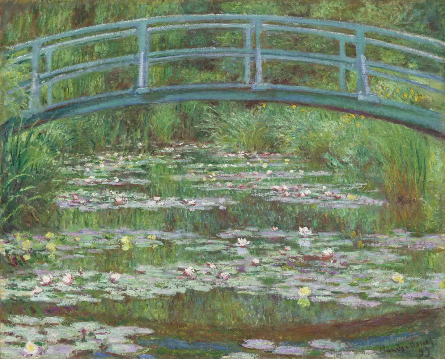
Claude Monet, The Japanese Footbridge (1899) · National Gallery of Art, Washington · public domain (CC0) · Wikimedia Commons — one of the 1899 versions (it is not confirmed that the source of the description in the text looked at this one)
The Fading Lens
The eye has a lens, just like a camera. The crystalline lens is a transparent lens located right behind the pupil that gathers incoming light and focuses it. As people age, this lens can become cloudy. Cataracts are an eye disease in which the crystalline lens becomes cloudy and light cannot pass through properly. Among cataracts, one type involves the center of the lens hardening into a yellow, and as it deepens, brown color, and dark brown cataracts break down the distinction between colors (PMID 26563659).
Cataracts do not change vision in just one way. An eye with cataracts can be imagined through three factors: the amount of light entering the eye, the composition of light by wavelength, and the degree to which light scatters inside the lens. To the viewer, these three come across as brightness, color, and haziness respectively, and a change in any of the three can affect vision (DOI: 10.1002/col.20503). Cataracts can make the world appear dim and hazy, and if it is a cataract where the crystalline lens turns yellow, colors can also look different.
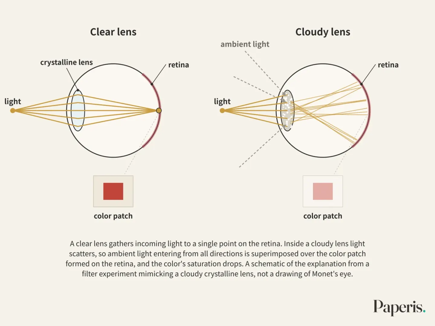
There is an experiment that isolated just the hazy feeling. In 2009, a color research team put goggles on participants fitted with hazy filters mimicking an older person's cloudy crystalline lens, and showed them red, yellow, green, and blue color patches while varying their size and the lighting (DOI: 10.1002/col.20503). This was an experiment using a simulation filter, not an experiment about Monet. When light scatters inside a cloudy lens, ambient light entering from all directions is superimposed over the color patch formed on the retina, causing that color to fade. Here, the word "fade" means that the saturation drops. Saturation refers to how vivid a color is.
The results varied greatly depending on size and light. At 300 lux, which is roughly the light of normal indoor lighting, a small color patch with a visual angle of 0.7 degrees hardly felt like it had any color. Under the same conditions, when the color patch was increased to a visual angle of 5.9 degrees, the perceived color increased rapidly. When the lighting was dimmed down to 10 lux, the degree of color fading decreased slightly, and the hazier the filter, the more the color faded (DOI: 10.1002/col.20503). The researchers explain that when a color patch is large, the color itself takes up the place of the light that was coming in right around it, reducing the degree of fading. In the filter experiment mimicking a cloudy crystalline lens, the colors beyond the cloudy lens did not always fade by the same amount, but varied depending on how large the color was and how bright the surroundings were.
The single sentence "cataracts change color" can simultaneously contain darkening, yellowing, and haziness, and in the simulation filter experiment, the results also differed depending on the viewing conditions.
What Happened to Monet's Eyes
Monet's cataracts began to develop in both eyes in his 60s. A two-page article published in a British general practice journal in 2015 notes this (DOI: 10.3399/bjgp15x684949). This matches a record in a 2003 article discussing the illnesses of Matisse and Monet, which states that cataracts were discovered in both eyes after a trip to Venice in 1908 when he was 68 (PMID 15147054).
In 1913, Monet went to London and consulted the German-born ophthalmologist Richard Liebreich. Liebreich had once been appointed professor of ophthalmology at St. Thomas's Hospital in London, and he was deeply interested in art himself, having published an article on the impact of eye disease on the paintings of the artists Turner and Mulready. He prescribed new glasses and recommended surgery on the right eye, but Monet refused (DOI: 10.3399/bjgp15x684949).
Around 1914 to 1915, remarks left by Monet are passed down like this. Colors no longer appeared with their former intensity, red began to look muddy, and his own paintings were becoming increasingly dark. To avoid choosing the wrong colors, Monet labeled his paint tubes and strictly maintained the order of paints on his palette. The glare of bright sunlight also troubled him, so he wore a large straw hat outdoors (DOI: 10.3399/bjgp15x684949). The labels on the paint tubes are the most concrete scene in this story. When his eyes could no longer be trusted with color, he had letters tell him the colors instead.
As Monet grew increasingly dispirited and painted less, his friend Georges Clemenceau, who had served as the Prime Minister of France and was also a physician, urged him to consider surgery. Monet feared the poor outcomes of cataract surgery experienced by his contemporaries, the artists Honoré Daumier and Mary Cassatt (DOI: 10.3399/bjgp15x684949).
Which eye was worse differs depending on the source. An article published in a university repository in 2008 records that Monet's right eye was officially legally blind while his left eye had 20/200 vision (DOI: 10.11575/prism/10378). 20/200 vision means that a person must approach to within 6 meters to recognize letters that a person with normal vision can recognize from about 60 meters away, which is 0.1 in our terms. On the other hand, the 2003 article records that he lost the vision in his left eye due to cataracts and that his right eye came to see colors abnormally (PMID 15147054). An article in JAMA from 1985 does not specify which eye, recording that after his lens became clouded and he was officially declared legally blind, he underwent surgery on his right eye (DOI: 10.1001/jama.1985.03360030084028). While the 1985 article and the 2003 article agree that the operated eye was the right eye, the 2008 article and the 2003 article conflict on which eye's vision was "lost."
Even after the right-eye surgery in 1923, the troubles did not end. Monet continued to paint after the surgery, but he suffered severe difficulties in balancing colors and struggled to adapt to the glasses worn after cataract surgery (DOI: 10.1001/jama.1985.03360030084028). The 2003 article records that a rare complication occurred after surgery where his field of vision appeared yellowish, which was partially corrected with Zeiss lenses (PMID 15147054). This story of the complication appears in only this single piece among the sources used in this article. Monet passed away on December 5, 1926, at the age of 86 (PMID 15147054).
Where did these records come from? The author of the 1985 JAMA article wrote the piece by reading a newly acquired bundle of letters sent by Monet to his eye surgeon at the French Ophthalmological Society, examining a pair of Monet's spectacles, and interviewing an optometrist and art historians who had worked alongside Monet's final ophthalmologist. The same author notes that the impact of eye difficulties on Monet's health and work has frequently been misunderstood (DOI: 10.1001/jama.1985.03360030084028). The 2008 article also notes the distance surrounding the surgery. A procedure that was routine for an ophthalmologist was unacceptable to Monet, and even though the two shared the same goal, they diverged sharply on the path to get there (DOI: 10.11575/prism/10378).
Let us place the words left by Monet side by side with the three elements from the previous section. On the color side, there are the statements that "colors do not appear with the same intensity as before" and "red looks muddy." The statement that "paintings are becoming increasingly dark" could mean that he perceived less brightness, or it could mean that he came to paint his canvases more darkly (DOI: 10.3399/bjgp15x684949). Regarding the hazy feeling, there are no statements by Monet remaining in the sources; instead, there is the glare of bright sunlight and the straw hat. This side-by-side arrangement merely shows which side Monet's discomfort leaned toward, but it does not tell us the exact extent of each of the three elements in his lens. Research connecting the result from a hazy goggle experiment—where large color areas appeared less faded—to Monet's thick brushstrokes is also absent from the sources of this article.
While multiple writings together convey Monet's cataracts and right-eye surgery, sources differ on which eye was worse. The fact that an eye disease "existed" and how profound that disease was in each eye are different questions, and reading the paintings in parallel requires an answer to the second question as well.
What Remained on the Canvas
Several medical papers state cause and effect directly. A 1985 article in “JAMA” writes that vision worsened by cataracts influenced Monet's late style, making him a painter of particular medical interest (DOI: 10.1001/jama.1985.03360030084028). A two-page article from 2015 writes that cataracts starting in his sixties eventually came to dramatically influence his work (DOI: 10.3399/bjgp15x684949). A 2008 article considers the way Monet's palette shifted over a ten-year period from 1912 to 1922 to reflect the progression of cataracts in both eyes (DOI: 10.11575/prism/10378). All three articles present that influence as the authors' judgment, and these abstracts contain no results from measuring the colors of specific paintings.
What those articles saw in the late paintings is this: the brushstrokes became thicker, and the paintings grew increasingly brownish, much like his cataracts (DOI: 10.3399/bjgp15x684949). "Like cataracts" is the phrasing of those authors. As his vision deteriorated, Monet found it harder to distinguish colors and began painting details less distinctly (DOI: 10.1001/jama.1985.03360030084028). An article transcribing a 2015 ophthalmology lecture writes that unfamiliar and intense use of color appears on Monet's late canvases before surgery (PMID 26563659), and a 2003 article writes that these eye changes can be recognized in his works (PMID 15147054). The 2008 article adds one more thing. It states that as he realized his vision was continuing to worsen, Monet's mindset and attitude toward his own painting also changed, and he could no longer trust the senses that had given him a lifetime of joy and success (DOI: 10.11575/prism/10378). In that article's reading, the change in mind also began from realizing that his vision was worsening.
Now we stand before two bridge paintings. On the left is the bridge from 1899. As a single-page article describes, greens, pale purples, and pinks reflect in the water to fill the canvas, and that article categorizes this painting as dating from before Monet's cataract problem (DOI: 10.4103/1995-705x.182650). On the right is the bridge from around 1920. What the preceding articles saw in the late paintings are thicker brushstrokes, colors leaning toward the brown side, less distinct details, and unfamiliar, intense color usage. Several years earlier, around 1914 to 1915, Monet remarked that reds looked muddy and his paintings had grown dark, and he labeled his paint tubes (DOI: 10.3399/bjgp15x684949). The facts placed between the two paintings connect chronologically in this way. However, none of the abstracts this article relies on as evidence contain results showing that specific colors of specific paintings were measured and attributed to cataracts.
Reading the color changes in the late paintings as connected to the progression of cataracts is an interpretation put forward in clear terms by several medical articles. What supports that interpretation in the abstracts available to this article is the chronological connection to recorded eye disease, not the results of measuring the colors of the paintings.
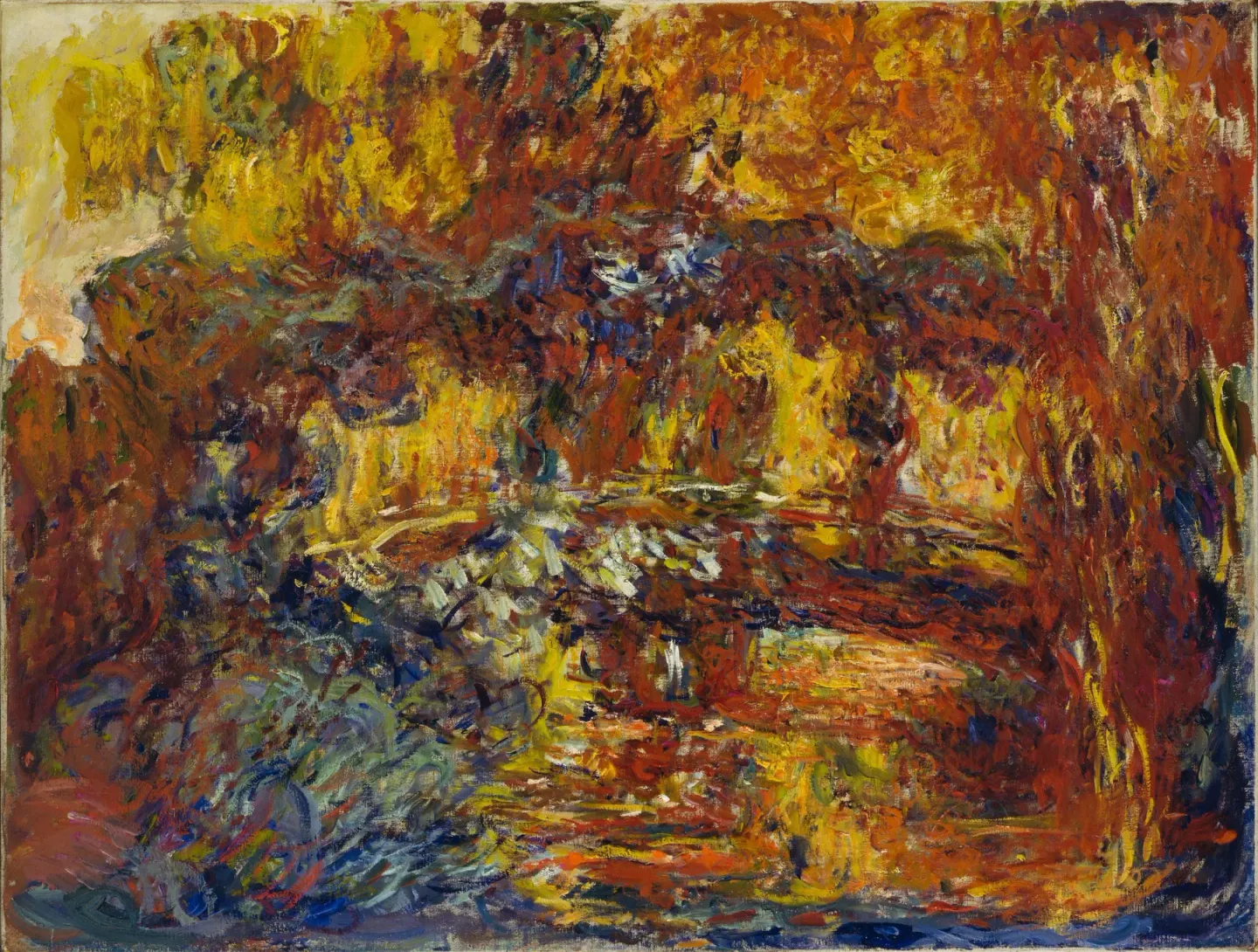
Claude Monet, The Japanese Footbridge (c. 1920–1922) · Museum of Modern Art, New York · public domain · Wikimedia Commons
Recreating Blurry Vision
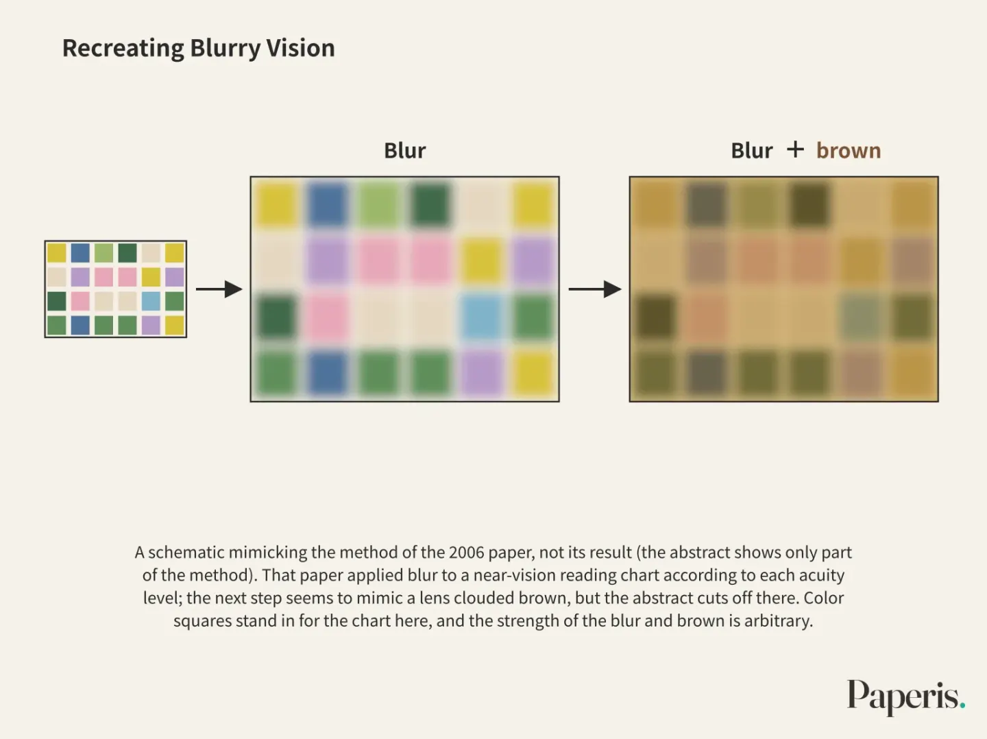
So, can we reproduce what Monet actually saw? A 2006 article published in an American ophthalmology journal attempted just that. The author reasoned that knowing how Monet and Degas actually saw the world and their own canvases in their later years would help us better understand what the two painters faced, and aimed to use medical knowledge and computer simulation to show the vision of the two painters and demonstrate how the two different diseases and different painting styles of the two men were related (DOI: 10.1001/archopht.124.12.1764). The first step of the method was to apply blur to a drawing of a near-vision reading chart. The intensity of the blur was determined according to which line could be read at each visual acuity level. The next step appears to lead into a process mimicking a brownishly blurred lens, but the abstract we have cuts off right at that word. Therefore, what the simulation showed cannot be conveyed in this article. Degas's disease, covered in the same paper, was a disease of the retina and thus different from Monet's cataract, and its results do not carry over to Monet.
What can be read from this method is what is required for recreation. How much to reduce the blur must be decided from visual acuity records, and if there is a brownish stage, the density of that color must also be decided. However, as we saw in the previous section, even which eye was how bad varies from evidence to evidence. In filter experiments mimicking a blurred lens, how much the color faded also varied depending on the size of the color patch and the lighting (DOI: 10.1002/col.20503). A recreated screen is only as accurate as the values put into it.
Just how much a single yellow lens changes color perception is being measured separately in modern ophthalmology. Absent in Monet's day, lenses are now newly inserted during cataract surgery. An intraocular lens is an artificial lens inserted in place of the clouded natural lens removed during cataract surgery. Among them, the one coated with a yellow tint is a lens that filters blue light (DOI: 10.1016/j.jcrs.2011.06.024). In a study from Brazil comparing 30 bilateral cataract patients 5 years after randomly inserting a yellow lens in one eye and a clear lens in the other, the color discrimination test results showed no statistically meaningful difference between the two eyes, and the contrast sensitivity under bright and dark conditions also showed no difference. The authors concluded that it is unclear whether the yellow lens has the advantage of protecting the center of the retina (DOI: 10.1016/j.jcrs.2011.06.024). A 2025 study from China looking at 54 patients and 108 eyes three months after surgery using a color arrangement test similarly found no meaningful difference in color perception overall between the yellow lens and the clear lens. However, in a subgroup comparing trifocal lenses, the tendency of color errors to scatter differed between the yellow lens and the clear lens (DOI: 10.1038/s41598-025-18259-5). Meanwhile, a 2007 Canadian study found that patients with yellow lenses needed more blue light than patients with clear lenses when judging brightness under bright conditions. The authors noted that the effect of this brightness reduction on daily life requires further study (DOI: 10.1016/j.jcrs.2007.03.024).
These studies did not measure Monet's eyes, and today's intraocular lenses are not the same condition as a lens clouded to a deep brown. In tests distinguishing colors, there was generally no difference, but in tests judging brightness, there was a difference. Even under the same label of "yellowish," results differed depending on the measurement method; that is the single takeaway this article takes from these studies.
Eye or Choice
Ophthalmology cannot explain art, but vision is a painter's tool, and its normality or abnormality can influence what a painter can do. A piece translating that 2015 ophthalmology lecture begins with this note. The same piece notes that diagnosing eye disease in a painter's work is dangerous because painters have the freedom to paint as they wish. As an example, the author writes that El Greco did not have astigmatism, Monet did not have myopia, and Turner did not have cataracts. However, it states that if record of an eye disease remains, its impact can be analyzed, and offers Monet as one such example. Dense brown cataracts break down color discrimination, and unfamiliar and strong color usage appears in Monet's late canvases before surgery (PMID 26563659). The lecture adds the caveat that it barely scratches the surface of the complexity of vision and art, yet concludes that understanding the eye and eye diseases opens several ways to look into art (PMID 26563659). Monet's cataract is a disease that remains in records, which differs in its starting point from a disease guessed backward from paintings.
Records do not provide all the answers. A 2003 piece dealing with painters' visual abnormalities notes that the world painted by painters often changes due to abnormalities in the visual pathway, that some painters deliberately used such distortions to create their own style, and that debates remain concerning older painters whose visual functions are not well preserved in historical records (PMID 12637042). A 2021 review surveying eye diseases of famous painters notes that a painting's style may have been deliberately created by the painter or influenced by eyesight, and that people have long wondered whether style was influenced by eyesight or not at all. However, this review itself sets as its goal to show how eye diseases could have influenced art creation over the past several centuries (PMID 35087973). A 2006 simulation piece also starts from the point that theoretical debates have continued over whether Monet's and Degas's deteriorated eyesight was a factor in their late styles or not (DOI: 10.1001/archopht.124.12.1764).
Gathering the evidence on Monet's side again, most abstracts collected by this article point toward the eye. There are Monet's own words that colors looked muddy and the labels on paint tubes (DOI: 10.3399/bjgp15x684949), the difficulty with color balance that continued even after surgery (DOI: 10.1001/jama.1985.03360030084028), and the changed mind and attitude as he realized his eyesight was failing (DOI: 10.11575/prism/10378). Articles arguing with evidence that Monet's late style was a deliberately chosen change regardless of his eyes are not among the evidence collected here. The possibility that the style might have been deliberately created remains only as a general theory (PMID 35087973), and some writings note that there are painters who deliberately used visual distortions as their style (PMID 12637042). This article reads this to mean that the eye and choice may not mutually exclude each other. How much of such choice was mixed into Monet's late style is not decided by the evidence in this article.
There is one variable left on the painting side as well. It is whether the colors of the late paintings we see today are the same as the colors Monet painted on that day. The fact that paints can change over time was covered in "Paint and Materials — The Same Powder, Different Doughs". Studies measuring the paint of Monet's late paintings and dealing with that change are not in the evidence of this article.
The reading that cataracts changed Monet's late colors is a retrospective interpretation put forward in clear terms by several medical writings, and whether the change in late style was solely due to the eyes is not decided by the evidence in this article.
Remaining Questions
Which eye was worse varies from one piece of evidence to another. What the 2006 simulation that recreated the blurry eye showed lies outside the abstract we have. Studies that measured specific colors in the late paintings and compared them to the changes in the eyes are also not among the evidence in this article. And whether the late bridge hanging on a museum wall today retains the exact colors Monet painted on that day cannot be answered by the evidence in this article either.
The bridge from 1899 and the bridge around 1920 are the same bridge over the same pond. That Monet's eyes became blurred in between is conveyed by multiple texts together, and that the colors of the paintings changed is shown by the paintings themselves. The line connecting the two is a line of interpretation clearly drawn by medical researchers, and whether all of the late-period colors can be explained by that single line cannot be determined by the evidence in this article.
Source Papers
- "The effect of cataracts and cataract surgery on Claude Monet" (2015) — DOI: 10.3399/bjgp15x684949 — A 2-page article in a British general practice journal. Bilateral senile cataracts in his 60s (ultimately having a dramatic impact on his work), the 1913 examination in London by Liebreich, new glasses, and the recommendation and refusal of surgery on his right eye, "the intensity of colors is not what it used to be, red is muddy, paintings are getting darker" in 1914–15, tube labels, palette order, straw hat, thicker brushstrokes and brown tones, Clemenceau's persuasion and fear of the surgical results of Daumier and Cassatt.
- "Ophthalmology and Art: Simulation of Monet's Cataracts and Degas' Retinal Disease" (2006) — DOI: 10.1001/archopht.124.12.1764 — Theoretical debate on how much vision was a factor in the late styles of Monet and Degas, contemporary reviews calling them "rough and crude," attempting to show fields of vision using medical knowledge and computer simulations and to demonstrate the connection between the two painters' different diseases and painting styles. The method involves progressive blurring on an eye chart to brown (green cuts off here, results not included).
- "Vision, eye disease, and art: 2015 Keeler Lecture." (2016) — PMID 26563659 — Ophthalmology cannot explain art, diagnosing eye diseases from artworks is dangerous (El Greco, Monet, Turner as examples), documented diseases can be analyzed, dense brown cataracts break down color discrimination, Monet's unfamiliar and strong colors on late-period canvases before surgery, concluding that understanding eyes and eye diseases opens a path to looking into art.
- "Cataracts and the Late Style of Monet's Paintings" (2008) — DOI: 10.11575/prism/10378 — An article hosted in a university repository (format unconfirmed). The argument that palette changes from 1912 to 1922 reflect the progression of bilateral cataracts, official blindness in the right eye and 20/200 in the left, changes in mind and attitude, and the mismatch between patient and doctor surrounding surgery.
- "Monet's Cataracts" (1985) — DOI: 10.1001/jama.1985.03360030084028 — The first painter called an Impressionist, vision worsened by cataracts affecting his late style, difficulty distinguishing colors and blurring of details, right eye surgery after official blindness, color balance and difficulty adapting to cataract glasses after surgery, the impact being frequently misunderstood, based on letters, glasses, optometry articles, and art historians housed in the French Society of Ophthalmology.
- "The influence of ophthalmological diseases on the vision quality of famous painters." (2021) — PMID 35087973 — Review. Style may be intentional or the effect of vision, and whether there is an effect has long been wondered.
- "Size effect of color patches for their color appearance with foggy goggles simulating cloudy crystalline lens of elderly people" (2009) — DOI: 10.1002/col.20503 — Three elements of a cataract eye (brightness, color, blur), experiment with cloudy filter goggles: color patches 0.7 degrees small are barely perceived as colored at 300 lux and increase rapidly when enlarged to 5.9 degrees, fading is slight at 10 lux, and fading is greater with more blur. Irrelevant to Monet.
- "[Influence of disease on Matisse and Monet painting]." (2003) — PMID 15147054 — Bilateral cataracts discovered after an 1908 trip to Venice, loss of left eye vision and abnormal color perception in the right eye, changes can be seen in the works, rare complication called xanthopsia (yellowish vision) after right eye surgery in 1923 and Zeiss lenses, death at age 86 on December 5, 1926.
- "Effects of blue light–filtering intraocular lenses on the macula, contrast sensitivity, and color vision after a long-term follow-up" (2011) — DOI: 10.1016/j.jcrs.2011.06.024 — Randomized 30 patients, 60 eyes, yellow (blue light-filtering) versus clear intraocular lenses for 5 years: no difference in color discrimination, contrast sensitivity, or macula, and the benefit of macular protection is unclear.
- "Luminance contrast with clear and yellow-tinted intraocular lenses" (2007) — DOI: 10.1016/j.jcrs.2007.03.024 — Patients with yellow intraocular lenses had lower luminance contrast values under bright conditions and required more blue light to judge brightness, and functional effects require further study.
- "Visual dysfunction in artists." (2003) — PMID 12637042 — Visual pathway abnormalities change an artist's expression, and some artists used this as a style, with debate surrounding old painters with poor records.
- "Evaluation of color perception in cataract patients bilateral implanted with presbyopia-correcting intraocular lenses" (2025) — DOI: 10.1038/s41598-025-18259-5 — 54 patients, 108 eyes, FM-100 3 months after surgery: overall no difference in color perception between yellow and clear intraocular lenses, with a difference in the S-index in the comparison of yellow versus clear within the trifocal lens subgroup.
- "Narrative and dialog" (2016) — DOI: 10.4103/1995-705x.182650 — A 1-page article. The 1899 "Water Lily Pond and Japanese Bridge" is a painting of green, pale purple, and pink, predating cataract problems.
Sources (13)
- The effect of cataracts and cataract surgery on Claude MonetDOI 10.3399/bjgp15x684949
- Ophthalmology and Art: Simulation of Monet's Cataracts and Degas' Retinal DiseaseDOI 10.1001/archopht.124.12.1764
- Vision, eye disease, and art: 2015 Keeler LecturePMID 26563659
- Cataracts and the Late Style of Monet's PaintingsDOI 10.11575/prism/10378
- Monet's CataractsDOI 10.1001/jama.1985.03360030084028
- The influence of ophthalmological diseases on the vision quality of famous paintersPMID 35087973
- Size effect of color patches for their color appearance with foggy goggles simulating cloudy crystalline lens of elderly peopleDOI 10.1002/col.20503
- [Influence of disease on Matisse and Monet painting]PMID 15147054
- Effects of blue light–filtering intraocular lenses on the macula, contrast sensitivity, and color vision after a long-term follow-upDOI 10.1016/j.jcrs.2011.06.024
- Luminance contrast with clear and yellow-tinted intraocular lensesDOI 10.1016/j.jcrs.2007.03.024
- Visual dysfunction in artistsPMID 12637042
- Evaluation of color perception in cataract patients bilateral implanted with presbyopia-correcting intraocular lensesDOI 10.1038/s41598-025-18259-5
- Narrative and dialogDOI 10.4103/1995-705x.182650